Guidelines for Bone Health

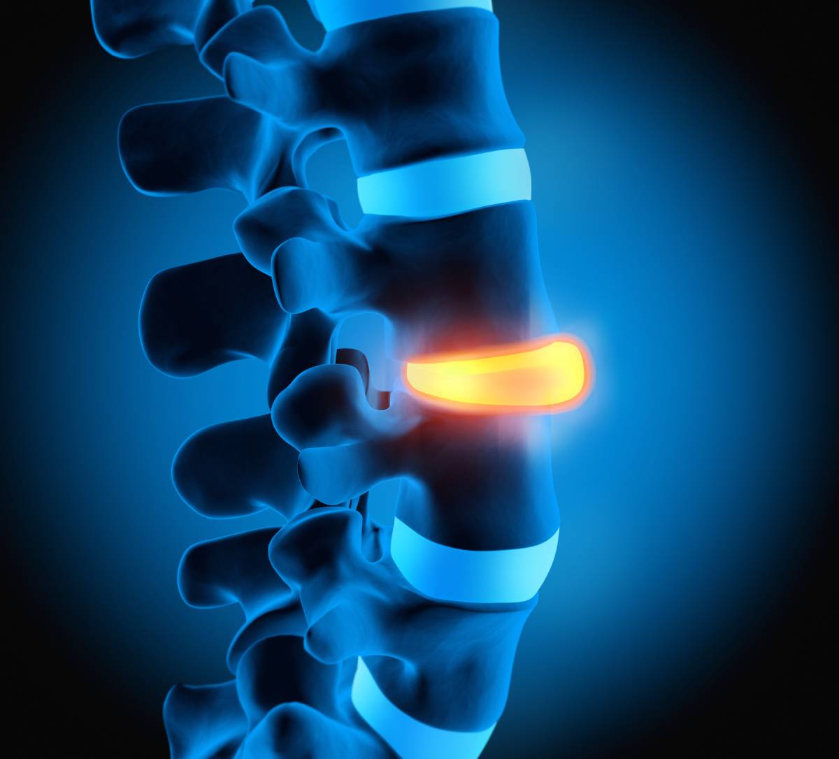
Deficiencies in bone health are common among elderly populations [1]. Associated musculoskeletal conditions, particularly osteoporosis, can result in significant physical disability and psychological impacts [1]. Fortunately, following guidelines for physical activity, nutrition, and pharmacological therapies can promote bone health and avoid the musculoskeletal problems that have become characteristic of old age.

Exercise is one of the most impactful ways through which people can improve their bone health [1]. Optimally, exercise should occur frequently during maturation to ensure that young people reach their peak bone mass and, accordingly, prevent or delay osteoporosis later in life [1]. Although exercise earlier in life can be very beneficial, it is impactful during middle and older age as well [1]. While the optimal form of exercise to promote bone health is not known, researchers recommend high-impact exercises that involve dynamic movements such as hopping or jumping [1, 2]. Alternatively, odd- or high-impact movements paired with resistance training can also be beneficial [2]. People who cannot engage in high-impact activities may benefit from certain yoga postures, lower-impact resistance training, or even walking [2]. Research indicates that habitually engaging in these forms of exercise for at least two or four short (30 minute or less) sessions each week is a good guidelines for maintaining or improving bone health [2].

Better nutrition is another lifestyle change that people can adopt to promote their bone health. Multiple studies have pointed to protein as highly influential for bone health; however, protein can be either detrimental or beneficial, depending on several factors [3]. Protein source is one hypothesized factor. Shams-White and colleagues investigated the differential effects of animal-based protein versus plant-based protein [3]. Their primary outcomes included lumbar spine (LS), femoral neck (FN), and total body bone mineral density (BMD) biomarkers [3]. Despite the two sources’ dissimilar amino acid profiles, research did not show a difference in benefits [3]. Another study conducted by the same researchers found that higher protein intake was more beneficial than lower intake for LS BMD, but otherwise, the two levels of intake were comparable in terms of FN, total hip, and total body BMD markers [4]. On another note, dairy products are a greater source of bone-beneficial proteins than any other food characteristic of the adult diet [5]. Indeed, in certain populations, calcium from dairy products appears to increase bone mineral density and improve total body bone mineral content when consumed over the long term [5].

When bone conditions have already progressed significantly, doctors often need to turn to pharmacological interventions [1]. Medications for bone conditions tend to be effective, but their benefits are often short-lived and associated with rare but drastic side effects [1]. For example, treatments such as bisphosphonates are known for their high success rates in treating patients who suffer from glucocorticoid-induced osteoporosis [6]. Unfortunately, bisphosphonates may not serve as an optimal long-term solution for certain patients, in which case additional management is required [6].

Ultimately, bone health can perhaps best be achieved by regularly following guidelines that have been established by thorough research, including a high-intensity impact workout regimen and consuming bone-nutrient-rich foods, such as dairy. When bone diseases have already progressed, pharmacological treatments can be highly effective, but their success may be limited to the short term. Therefore, maintaining healthy nutritional and exercise habits is of the utmost importance to avoid persistent bone problems.

References 

[1] L. Santos, K. J. Elliott-Sale, and C. Sale, “Exercise and bone health across the lifespan,” Biogerontology, vol. 18, no. 6, p. 931-946, October 2017. [Online]. Available: https://doi.org/10.1007/s10522-017-9732-6

[2] K. L. Troy et al., “Exercise Early and Often: Effects of Physical Activity and Exercise on Women’s Bone Health,” International Journal of Environmental Research and Public Health, vol. 15, no. 5, p. 1-17, April 2018. [Online]. Available: https://doi.org/10.3390/ijerph15050878

[3] M. M. Shams-White et al., “Animal versus plant protein and adult bone health: A systematic review and meta-analysis from the National Osteoporosis Foundation,” PLoS One, vol. 13, no. 2, p. 1-24, February 2018. [Online]. Available: https://doi.org/10.1371/journal.pone.0192459

[4] M. M. Shams-White et al., “Dietary protein and bone health: a systematic review and meta-analysis from the National Osteoporosis Foundation,” The America Journal of Clinical Nutrition, vol. 105, no. 6, p. 1528-1543, June 2017. [Online]. Available: https://doi.org/10.3945/ajcn.116.145110

[5] E. G. H. M. van den Heuvel and J. M. J. M. Steijns, “Dairy products and bone health: how strong is the scientific evidence?,” Nutrition Research Reviews, vol. 31, p. 164-178, March 2018. [Online]. Available: https://doi.org/10.1017/S095442241800001X

[6] A. Caplan et al., “Prevention and management of glucocorticoid-induced side effects: A comprehensive review: A review of glucocorticoid pharmacology and bone health,” Journal of American Academy of Dermatology, vol. 76, no. 1, p. 1-9, January 2017. [Online]. Available: https://doi.org/10.1016/j.jaad.2016.01.062

Chiropractic Care for Newborn Torticollis

Infant torticollis manifests when the sternocleidomastoid muscle, connecting the breastbone and collarbone to the skull, is shortened as a result of musculoskeletal or non-musculoskeletal factors 1, either at birth in the form of congenital torticollis, or later on in development in the form of acquired torticollis. The worldwide incidence of congenital torticollis varies from 0.3% to 1.9 %, with some studies pointing to a prevalence of 1 in 250 newborns, representing the third most frequent congenital orthopedic anomaly 2. An early diagnosis of newborn torticollis facilitates its swift noninvasive correction, such as through chiropractic care, minimizing any long-term complications. With proper treatment, 90% to 95% of children improve during their first year of life, and 97% of patients improve if treatment is begun prior to six months of age 3.

There are several methods of treating congenital torticollis. Physical and chiropractic therapy and repositioning are recommended as a first line treatment option. Such initial treatment focuses on passive range stretching and thorough patient follow ups, including manual flexion, extension, and lateral rotation stretches. Good stabilization and correct hand positioning are key, although stretching methods and positions are highly patient-centric 4.

A 2015 case study demonstrated that chiropractic management of infantile torticollis fully resolved the torticollis and associated abnormal fixation of the right eye 5. Chiropractic treatment consisted of cervical spine manipulative therapy alongside neck muscle massage and stretching; all symptoms resolved within 4 weeks.

Since, chiropractic manipulation under anesthesia has been developed as a novel therapeutic method. In a 2018 retrospective case series, 6 infants aged 4.5- to 15-months-old previously diagnosed with newborn torticollis, after failing to respond to conservative therapy options, were treated by a chiropractic doctor. All 6 infants, who were found to have arthrogenic newborn torticollis, responded well to manipulation under anesthesia 6, experiencing a significantly improved cervical range of motion. Manipulation under anesthesia may represent a therapeutic option for complex arthrogenic torticollis cases failing to respond to other adjunctive therapies.

Most recently, a 2020 case report documented the full resolution of an infant’s congenital torticollis using a multidisciplinary chiropractic approach. The treatment consisted of a gentle manipulation of the occipito-atlantal spinal region, soft tissue therapy of the sternocleidomastoid muscle, and cranial manipulation alongside a number of home exercises and a referral to a physiotherapist 7. Since, a 2020 systematic review assessing the effectiveness and safety of conservative interventions for torticollis at all ages corroborated that practitioner-led stretching and repositioning practices represented low risk, inexpensive interventions with tremendous potential for treating newborn torticollis 8. Finally, alongside these interventions, both Kinesio Taping and helmet therapy, primarily for infants with moderate to severe and persisting asymmetry, provide additional chiropractic care tools for the treatment of newborn torticollis 4

Research has highlighted the positive outcomes of a multidisciplinary approach to correcting pediatric torticollis. Parents should remain well-educated as regards the condition and its prognosis, alongside proactively participating in its management. In concert, healthcare providers should remain aware of the association between congenital torticollis and its impact on a child’s gross motor developmental milestones. In the end, prompt, patient-adapted therapy from a collaborative team will remain key to sustainable clinical outcomes 3.  

References

1.        Haugen, E. B., Benth, J. Š. & Nakstad, B. Manual therapy in infantile torticollis: A randomized, controlled pilot study. Acta Paediatr. Int. J. Paediatr. (2011). doi:10.1111/j.1651-2227.2011.02145.x

2.        Congenital torticollis – Physiopedia. Available at: https://www.physio-pedia.com/Congenital_torticollis.

3.        Gundrathi, J., Cunha, B. & Mendez, M. D. Congenital Torticollis. Anaesthesia, Pain Intensive Care 20, 376 (2021).

4.        Öhman, A. M. The Immediate Effect of Kinesiology Taping on Muscular Imbalance for Infants With Congenital Muscular Torticollis. PM R (2012). doi:10.1016/j.pmrj.2012.04.006

5.        Hobaek Siegenthaler, M. Chiropractic management of infantile torticollis with associated abnormal fixation of one eye: A case report. J. Chiropr. Med. (2015). doi:10.1016/j.jcm.2014.12.003

6.        Paravicini, I. Manipulation Under Anesthesia in Infants With Arthrogenic Newborn Torticollis: A Retrospective Case Series. J. Chiropr. Med. (2018). doi:10.1016/j.jcm.2018.04.004

7.        Lacerte, F. D. D. Positive outcome from a chiropractic multidisciplinary approach for congenital torticollis in an infant three-weeks of age: A case report. J. Clin. Chiropr. Pediatr. (2020).

8.        Ellwood, J., Draper-Rodi, J., Carnes, D. & Carnes, D. The effectiveness and safety of conservative interventions for positional plagiocephaly and congenital muscular torticollis: A synthesis of systematic reviews and guidance. Chiropractic and Manual Therapies (2020). doi:10.1186/s12998-020-00321-w

Bulging Disc vs. Herniated Disc

In light of an increasingly sedentary society, back pain is growing increasingly common among the general population (1), resulting from a number of ailments – of which bulging or herniated discs are quite frequent.

A bulging disc is formed when the soft cushion of tissue between vertebrae pushes outward, resulting in consistent pressure. In contrast, a herniated disc is formed when the outer covering of the disc is torn, causing the innermost nucleus pulposus to leak into the spinal canal. Both bulging and herniated discs result in significant pain and tenderness, although bulging discs tend to generate a radiating sensation of dull pain. In more severe cases, bulging or herniated discs may impinge traversing nerves as these exit the neural foramen, causing nerve pain, or even directly compress the spinal cord contained within the spinal canal. Both most frequently affect the cervical and thoracic spinal regions (2).

Clinically considered degenerative, a bulging disc presents as a progressive, gradual onset of symptoms. Symptoms can include pain in the buttocks, legs, or back, preventing a patient from walking smoothly. Bulging discs tend to affect multiple discs and may generate other disc degeneration-related issues, such as lumbar stenosis, or a narrowing of the spinal canal (3). Short-term treatment includes the administration of anti-inflammatory medications, especially during acute flare-ups. Steroids may also be administered in the case of significant nerve pain. Thereafter, long-term treatment involves a self-directed exercise program which is begun with a physical therapist prior to being continued at home. In the case of lumber stenosis, lumbar decompression surgery – decompressing the spinal canal such that the nerves are no longer compressed to alleviate the pain – may be warranted.

Herniated discs generally occur abruptly, as a result of an acute injury, and affect only one individual nerve root (3). If symptoms have lasted less than 6 weeks and patients have not experienced any nerve damage, anti-inflammatory medications, physical therapy or chiropractic treatment, and steroid injections can help address symptoms over a 6- to 12-week period. Most modern chiropractors use a variety of effective treatment approaches, including gentle soft tissue therapies, graded mobilization, and intermittent traction. If these less invasive forms of treatment are not sufficiently effective, an outpatient surgery in the form of a microdiscectomy may quickly alleviate the pain by relieving the pressure on the affected nerve root. Certain extreme cases of disc herniation require tailored treatment. For example, lumber disc herniation into the dural space, a rare phenomenon associated with degenerative lumbar lesions among the elderly, requires immediate surgery, yielding generally positive outcomes (4). It should not go unnoticed that, interestingly, a recent study showed that common terms used in lumbar spine imaging reports are poorly understood by the general population and may contribute to the burden of lower back pain (5).

Overall, non-surgical treatments such as chiropractic care are effective and are the preferred first line treatment for a bulging or herniated disc. Alongside lucid and thorough patient education, patient-tailored treatments of varying degrees of invasiveness help patients rapidly resume their normal activities in a safe and sustainable fashion.

References 

1. Hanna, F. et al. The relationship between sedentary behavior, back pain, and psychosocial correlates among university employees. Front. Public Heal. (2019). doi:10.3389/fpubh.2019.00080 

2. Bulging Discs vs. Herniated Discs: Understanding the Difference – Chiropractic BioPhysics. Available at: https://idealspine.com/bulging-discs-vs-herniated-discs-understanding-the-difference/. 

3. Bulging Disc Vs. Herniated Disc: What’s The Difference? – Penn Medicine. Available at: https://www.pennmedicine.org/updates/blogs/musculoskeletal-and-rheumatology/2018/november/bulging-disc-vs-herniated-disc. 

4. Ge, C. Y. et al. Intradural lumbar disc herniation: A case report and literature review. Clin. Interv. Aging (2019). doi:10.2147/CIA.S228717 

5. Farmer, C. et al. 53 Consumer understanding of terms used in imaging reports requested for low back pain. in (2019). doi:10.1136/bmjebm-2019-pod.66 

The Role of Kinesiotaping in Chiropractic Care

            Kinesiotaping is a popular therapeutic approach in the overlap between physical therapy, sports medicine, and chiropractic care. Flexible pieces of kinesiotape are placed strategically around the affected area in order to guide muscular movement, improve circulation, and relieve pain. The tape is both waterproof and durable; it can therefore be worn during intense physical exercise as well as during rest. Often, tape does not need to be replaced by a practitioner for several days.

            Given the flexibility of its applications as well as its multidimensional benefits, kinesiotaping has shown particular promise for the adjustment of posture and alignment during motion, particularly when it comes to foot pronation. Indeed, several studies have suggested that kinesiotaping may be a promising treatment for excessive pronation.1,2,3 In order to verify these findings, a 2020 study by physical therapists Senem Guner and Serap Alsancak aimed to characterize the benefits of kinesiotaping as a therapeutic intervention for foot pronation.4 A total of twenty participants (split equally by gender) with a current diagnosis of foot pronation were included in the study.

            In order to optimize kinesiotape efficacy, the authors projected a laser line onto each participant as they stood, barefoot, with one foot on a force plate, thus illuminating the joint load carrying line. A practitioner then applied kinesiotape in a corrective fashion. Assessment of the joint load carrying line was done prior to kinesiotape application, immediately after, and then at 24-48 hours following application. A ruler was placed at the level of the joint line tangentially to the patella and ankle joint in order to quantify deviation of the joint load carrying line.

            Interestingly, Guner and Alsancak found that kinesiotape corrected neither the weight bearing nor the joint load carrying line in any significant way. While improvement was noted in lateral knee joint movement at the time of application of the kinesiotape, this change did not persist into the 24-48-hour mark. This finding suggests that kinesiotape may experience waning efficacy with continued wear, perhaps due to stretching and/or loosened adhesion, and that the tape should be reapplied more frequently to maximize effectiveness.

            While these findings are somewhat contradictory to those of previous studies, it is important to note a few pertinent factors which may have contributed. Firstly, the authors’ sample size was small and not particularly diverse in age (average age 19.7 ± 1.2 years); there is therefore ample opportunity for future studies to both replicate and expand on these findings. Moreover, other pertinent values which were assessed in other similar studies – such as changes in peak plantar pressure – were not assessed by Guner and Alsancak. A more multidimensional study may have revealed a more nuanced effect.

The authors do note that kinesiotaping still may be useful for amplifying sensory input and reduction of perceived discomfort. Furthermore, short-term use of kinesiotape may also prove more effective when paired with orthotic footwear. However, the authors findings do indicate the need for continued research.

References

1. Franettovich, M. M., Murley, G. S., David, B. S., & Bird, A. R. (2012). A comparison of augmented low-Dye taping and ankle bracing on lower limb muscle activity during walking in adults with flat-arched foot posture. Journal of science and medicine in sport15(1), 8–13. https://doi.org/10.1016/j.jsams.2011.05.009

2. Franettovich, M., Chapman, A., & Vicenzino, B. (2008). Tape that increases medial longitudinal arch height also reduces leg muscle activity: a preliminary study. Medicine and science in sports and exercise40(4), 593–600. https://doi.org/10.1249/MSS.0b013e318162134f

3. Lange, B., Chipchase, L., & Evans, A. (2004). The effect of low-Dye taping on plantar pressures, during gait, in subjects with navicular drop exceeding 10 mm. The Journal of orthopaedic and sports physical therapy34(4), 201–209. https://doi.org/10.2519/jospt.2004.34.4.201

4. Guner, S., & Alsancak, S. (2020). Kinesiotaping Techniques to Alter Static Load in Patients With Foot Pronation. Journal of chiropractic medicine19(3), 175–180. https://doi.org/10.1016/j.jcm.2019.12.004

Fall Injuries and Prevention in Older Adults

In the United States, falls and fall-related injuries occur frequently [1]. Among the population aged 65 years and older, about 25% of people experience a fall annually [1]. While these falls do not always result in serious injury or fatality, they are still incredibly costly and risky overall [1]. Each year, falls and related injuries account for at least $50 billion in medical costs, 2.8 million visits to emergency departments, and 27,000 deaths [1]. Falls are also a major contributor to hospital readmission rate [2]. Considering that fewer than 25% of fall-related injuries are accurately reported, these numbers are likely an underestimate [3]. Accordingly, it is essential to know the risk factors associated with fall injuries, as well as the ideal prevention techniques necessary to reduce the ubiquity of this occurrence, especially among older adults.

The risk factors for fall injuries are manifold. They include environmental factors, such as uneven floors, missing hand-rails, and poor lighting [4]. Chronic conditions that affect mobility and/or cognition, such as Parkinson’s Disease, stroke, diabetes, and arthritis, can increase an older adult’s likelihood experiencing a fall and subsequent injuries [5]. Additionally, acute illnesses like pneumonia and urinary tract infections augment the likelihood that a person will fall [5]. Physiologically, visual and hearing impairments, sarcopenia, low body mass index, and postural hypertension can also heighten the risk of falling among older adults [5].

Over recent years, further research has revealed more unintuitive risk factors. For instance, Lohman, Fairchild, and Merchant identified a possible association between antidepressant medication use and falls and fall injuries [1]. In their study of 3,565 community-dwelling older adults (aged 65 years or older), antidepressant medication accounted for about 19% and 18% of the association between major depressive disorder and falls and fall injuries respectively [1]. Other psychoactive medications have also been associated with greater fall risk [5].

Fall prevention is an essential element in the care of older adults, particularly those with several risk factors. However, it can be difficult to successfully navigate, with patients, payers, providers, and the overall health care system often impairing the quality of care available to older adults. Furthermore, strategies that appear as though they would be beneficial can fail to reduce fall rates. One such example is multifactorial interventions, such as the one studied by Bhasin in 2020 [6]. Despite providing patients with individualized plans and risk assessments, all administered by specialized nurses, the rate of fall injury did not diminish compared to standard care [6].

This is not to say that all multi-pronged approaches are unsuccessful: Kruschke and Butcher recommended an evidence-based ten-step protocol including fall, gait, and balance screening [4]. Unfortunately, those experimenters did not provide data on the success of their protocol [4]. However, other studies indicate that some multifactorial intervention programs can reduce the rate at which older adults fall [5]. Along with incorporating various screenings and risk factor-specific guidelines, medical practitioners should also advise their patients to regularly engage in physical activities, such as Tai Chi [5]. As seen with antidepressants, doctors should prescribe psychoactive medications with potential effects on fall risk in mind.

Because of the multitude of factors that can influence patients’ likelihood of falling, fall prevention is difficult to navigate. Regardless, analyzing risk factors, adjusting medications, screening appropriately, and promoting regular exercise can help reduce fall risk and rate.

References 

[1] M. C. Lohman, A. J. Fairchild, and A. T. Merchant, “Antidepressant Use Partially Mediates the Association Between Depression and Risk of Falls and Fall Injuries Among Older Adults,” The Journals of Gerontology: Series A, vol. 76, no. 9, p. e171-e178, October 2020. [Online]. Available: https://doi.org/10.1093/gerona/glaa253.  

[2] G. J. Hoffman et al., “Posthospital Fall Injuries and 30-Day Readmissions in Adults 65 Years and Older,” JAMA Network Open, vol. 2, no. 5, p. 1-12, May 2019. [Online]. Available: https://doi.org/10.1001/jamanetworkopen.2019.4276

[3] G. J. Hoffman et al., “Underreporting of Fall Injuries of Older Adults: Implications for Wellness Visit Fall Risk Screening,” Journal of the American Geriatrics Society, vol. 66, no. 6, p. 1195-1200, April 2018. [Online]. Available: https://doi.org/10.1111/jgs.15360

[4] C. Kruschke and H. K. Butcher, “Evidence-Based Practice Guideline: Fall Prevention for Older Adults,” Journal of Gerontological Nursing, vol. 43, no. 11, p. 15-21, October 2017. [Online]. Available: https://doi.org/10.3928/00989134-20171016-01

[5] R. E. Taylor-Piliae and R. Peterson, “Clinical and Community Strategies to Prevent Falls and Fall-Related Injuries Among Community-Dwelling Older Adults,” Nursing Clinics, vol. 52, no. 3, p. 489-497, September 2017. [Online]. Available: https://doi.org/10.1016/j.cnur.2017.04.004

[6] S. Bhasin, “A Randomized Trial of a Multifactorial Strategy to Prevent Serious Fall Injuries,” The New England Journal of Medicine, vol. 383, p. 129-140, July 2020. [Online]. Available: https://doi.org/10.1093/gerona/glaa253.  

Iron: Dietary Sources and Physiological Role

Iron is a metallic element that participates in chemical reactions important for biological processes. The physiological role of iron is as a component of heme and in other protein functional groups (Dev and Babitt, 2017). It is generally found in two different ionic forms: Fe2+, the reduced form and Fe3+, the oxidized form (Dev and Babitt, 2017). In excess, iron in the Fe2+ form can lead to the production of toxic free radicals that can damage cell structures; it is thus highly regulated in the body (Vogt et al, 2021).

Iron is brought into the body through the diet. The estimated average requirement for iron intake is 5 to 11.4 mg/day (Lim et al, 2013). Meat, seafood, legumes and fortified cereals are all significant dietary sources of iron (Lim et al, 2013). Although plant-derived foods contain slightly more iron on average than animal-derived foods, iron from heme in animal tissues is more bioavailable — more able to be absorbed and requiring less energy to be useful in its physiological role (Lim et al, 2013). Phytates (a form of phosphate storage in seeds and grains) and polyphenols (antioxidants found in tea and coffee) reduce the bioavailability of iron. Vitamin C, on the other hand, increases bioavailability (Lim et al, 2013).

In the digestive tract, iron is taken up by the cells of the duodenum. It must be in the Fe2+ form for this to happen, so iron that is in the Fe3+ form is converted by the cytochrome DCYTB (Vogt et al, 2021). Fe2+ ions enter duodenal cells through the transport protein DMT1. The iron can then be sent to the bloodstream by the transport protein ferroportin on the other side of the cell. It is immediately converted back to Fe3+ by the proteins ceruloplasmin and hephaestin and binds to transferrin, a carrier protein in the bloodstream (Vogt et al, 2021).

The most common physiological role of iron in the body is for hemoglobin in red blood cells, also known as erythrocytes (Vogt et al, 2021). Erythroid precursor cells have transferrin receptor 1. When transferrin binds to the receptor, the complex is internalized by the cell. The Fe3+ that was bound to the transferrin is released and is turned back into Fe2+ by protein STEAP3. The iron is finally available to be incorporated into heme (Vogt et al, 2021). A similar process occurs in the muscle cells for the production of myoglobin, which allows muscles to maintain their own small stores of oxygen. Cells can also store iron that they intake with the intracellular protein ferritin. Cells are able to regulate their iron storage through how many ferritin proteins and transferrin receptors they make (Vogt et al, 2021).

Systemically, iron levels are regulated by the liver. The liver produces the hormone hepcidin, which causes ferroportin to be destroyed. Doing this prevents excess iron from entering circulation (Dev and Babitt, 2017). Additionally, immune system macrophages contribute to iron regulation by recycling iron stores. Once the macrophages have cleaned up an old or damaged red blood cell, they can remove the iron from the hemoglobin and use ferroportin to transport it back into the bloodstream (Dev and Babitt, 2017).

Problems with iron homeostasis can lead to health complications. One example is anemia, which is a problem with red blood cells’ ability to carry oxygen. Iron deficiency anemia can occur when there is not enough dietary iron available; excessive iron loss, such as bleeding or chronic kidney disease; genetic issues with any of the proteins mentioned previously; or chronic immune conditions that cause an overproduction of hepcidin (Dev and Babitt, 2017). On the other hand, iron overload can cause highly reactive forms of iron to be taken up by organs such as the heart, liver and endocrine tissues and cause lasting damage and dysfunction (Dev and Babitt, 2017).

References

Dev S, Babitt JL. Overview of iron metabolism in health and disease. Hemodial Int. 2017;21 Suppl 1(Suppl 1):S6-S20. doi:10.1111/hdi.12542

Lim KH, Riddell LJ, Nowson CA, Booth AO, Szymlek-Gay EA. Iron and zinc nutrition in the economically-developed world: a review. Nutrients. 2013;5(8):3184-3211. Published 2013 Aug 13. doi:10.3390/nu5083184

Vogt AS, Arsiwala T, Mohsen M, Vogel M, Manolova V, Bachmann MF. On Iron Metabolism and Its Regulation. Int J Mol Sci. 2021;22(9):4591. Published 2021 Apr 27. doi:10.3390/ijms22094591

Omega-3 Fatty Acids for Joint Health

Omega-3 fatty acids are unsaturated fatty acids found in foods such as fish and flaxseed and in dietary supplements such as fish oil.[i] They are an essential nutrient, meaning that the body cannot synthesize them naturally but must derive them from food.[ii] Omega-3 fatty acids play an important role in many physiological functions, including joint health.

The three main omega-3 fatty acids are alpha-linolenic acid (ALA), eicosapentaenoic acid (EPA), and docosahexaenoic acid (DHA); while EPA and DHA can be synthesized from ALA, the conversion efficiency is low and thus all three are considered “essential.”[iii] Omega-3 fats are involved in several important cellular processes, including the production of hormones that regulate blood clotting and inflammation and the formation of cell membranes.2      

Omega-3 fatty acids have long been highlighted by the American Heart Association for their ability to reduce the incidence of cardiovascular events like heart attack and stroke.[iv] They are also integral for the function of the nervous system and the retina.[v] Many of these health benefits first came to light in the 1980s, when studies showed that the Greenland Inuit, whose diets are rich in seafood, have a dramatically lower incidence of cardiac and other health disorders than Western Europeans.[vi]

Additionally, omega-3 fatty acids have been shown to be an effective addition to boost joint health in treatment regimens for musculoskeletal disorders, particularly rheumatoid arthritis (RA). This is primarily due to the anti-inflammatory action of omega-3 fatty acids. RA occurs when the immune system attacks healthy cells in the joints, causing pain and inflammation.[vii] Many studies have used fish oil to demonstrate the anti-inflammatory effects of omega-3 fatty acids. For example, Espersen et al. showed that the blood plasma level of interleukin-1 beta, an inflammatory cytokine, decreases after fish oil consumption.[viii] Other mechanisms of action for the anti-inflammatory properties of omega-3 fatty acids may include the inhibition of T-cell proliferation and the reduction of antigen presentation.[ix]

A 2013 study published in the British Journal of Clinical Pharmacology argues convincingly for anti-inflammatory effects of omega-3 fatty acids. The paper[x] touches on the effect omega-3 fatty acids have on the chemotaxis, or movement, of white blood cells, which contribute to the development of arthritis when they attack the lining of the joint capsule.[xi] White blood cells are attracted to inflammatory sites by “chemo-attractants” such as the chemical LTB4.10 The author cites several studies which support the notion that fish oil supplements, and ultimately, the omega-3 fatty acids they contain, can restrict the chemotaxis of white blood cells toward chemoattractants. The mechanism of action has not yet been deduced, but the paper hypothesizes that it may involve the antagonism of receptors for chemo-attractants. The review also discusses the amount of omega-3 fatty acids needed to elicit their anti-inflammatory effects. It notes that typical consumption of “marine” omega-3 fatty acids (those coming from fish or seafood) is in the tens or low hundreds of milligrams per day.[xii] However, as Calder points out, multiple studies show that much larger amounts of omega-3 fatty acids are needed to reduce inflammation in those with chronic conditions: a study of rheumatoid arthritis showed 3.5g of EPA to be necessary to significantly reduce inflammation.[xiii] The review therefore suggests that those with inflammatory conditions take omega-3 supplements that far surpass what one would receive from a natural diet.


References

[i] “Office of Dietary Supplements – Omega-3 Fatty Acids.” NIH Office of Dietary Supplements, U.S. Department of Health and Human Services, 4 Aug. 2021, ods.od.nih.gov/factsheets/Omega3FattyAcids-Consumer/.

[ii] “Omega-3 Fatty Acids: An Essential Contribution.” The Nutrition Source, Harvard T.H. Chan School of Public Health, 22 May 2019, www.hsph.harvard.edu/nutritionsource/what-should-you-eat/fats-and-cholesterol/types-of-fat/omega-3-fats/.

[iii] “Essential Fatty Acids.” Linus Pauling Institute, Oregon State University, 1 Jan. 2021, lpi.oregonstate.edu/mic/other-nutrients/essential-fatty-acids.

[iv] “Fish and Omega-3 Fatty Acids.” www.heart.org, American Heart Association, www.heart.org/en/healthy-living/healthy-eating/eat-smart/fats/fish-and-omega-3-fatty-acids.

[v] Querques, G., et al. “Retina and Omega-3.” Journal of Nutrition and Metabolism, vol. 2011, 2011, pp. 1–12., doi:10.1155/2011/748361.

[vi] Kromann, N., and A. Green. “Epidemiological Studies in The Upernavik District, Greenland.” Acta Medica Scandinavica, vol. 208, no. 1-6, 2009, pp. 401–406., doi:10.1111/j.0954-6820.1980.tb01221.x.

[vii] “Rheumatoid Arthritis (Ra).” Centers for Disease Control and Prevention, Centers for Disease Control and Prevention, 27 July 2020, www.cdc.gov/arthritis/basics/rheumatoid-arthritis.html.

[viii] Espersen, G. T., et al. “Decreased Interleukin-1 Beta Levels in Plasma from Rheumatoid Arthritis Patients after Dietary Supplementation with n-3 Polyunsaturated Fatty Acids.” Clinical Rheumatology, vol. 11, no. 3, 1992, pp. 393–395., doi:10.1007/bf02207200.

[ix] Kostoglou-Athanassiou, I., et al. “The Effect of Omega-3 Fatty Acids On Rheumatoid Arthritis.” Mediterranean Journal of Rheumatology, vol. 31, no. 2, 2020, p. 190., doi:10.31138/mjr.31.2.190.

[x] Calder, P. C. “Omega-3 Polyunsaturated Fatty Acids and Inflammatory Processes: Nutrition or Pharmacology?” British Journal of Clinical Pharmacology, vol. 75, no. 3, 2013, pp. 645–662., doi:10.1111/j.1365-2125.2012.04374.x.

[xi] “Arthritis.” Mayo Clinic, Mayo Foundation for Medical Education and Research, 19 July 2019, www.mayoclinic.org/diseases-conditions/arthritis/symptoms-causes/syc-20350772.

[xii] Meyer, B. J., et al. “Dietary Intakes and Food Sources of Omega-6 and Omega-3 Polyunsaturated Fatty Acids.” Lipids, vol. 38, no. 4, 2003, pp. 391–398., doi:10.1007/s11745-003-1074-0.

[xiii] Calder, P. C. “Session 3: JOINT Nutrition Society and IRISH Nutrition and Dietetic Institute Symposium ON ‘Nutrition and Autoimmune Disease’ Pufa, Inflammatory Processes and Rheumatoid Arthritis.” Proceedings of the Nutrition Society, vol. 67, no. 4, 2008, pp. 409–418., doi:10.1017/s0029665108008690.

Chiropractic Adjustment: Manual Adjustment and Drop Table

Given the likelihood that 84% of the population will experience back pain in their lifetimes, there is a high demand for treatments to address this condition [1]. Due to its ability to assuage back pain, along with neck pain and headaches, chiropractic adjustment is one such option [2]. Generally, adjustment procedures on the back consist of a doctor of chiropractic applying a controlled force to the patient’s spinal joints, either with their hands or an instrument [2]. If successful, the patient will experience diminished or eliminated stiffness and soreness [2]. There are many methods for performing a chiropractic adjustment, including manual adjustment and drop table [2].

Manual adjustment involves chiropractors using their hands to apply quick, low-amplitude thrusts on the patient’s spinal joints [3]. It is one of the most common forms of chiropractic adjustment, owing both to the general population’s familiarity with the technique and doctors’ tendency to be comfortable administering the method [3]. Manual adjustment is often successful when treating joint soreness, pain, or stiffness [3]. More specifically, it may be an ideal technique to use when other methods, such as laser or infrared technology, have failed to relieve patient’s conditions [3].

There is no single ideal candidate for manual chiropractic adjustment: anyone who suffers from back, neck, or other joint pain, stiffness, or soreness can benefit from this form of treatment [3]. It can relieve subluxations caused by toxins, trauma, or mental difficulties [3]. Overall, manual adjustment is a highly versatile chiropractic technique.

An alternate form of chiropractic adjustment is the drop table method. For drop table adjustments, the chiropractor uses the force of gravity, administered via a controlled drop in a segment of the treatment table, to settle the patient’s spine into place [4]. It requires considerably less applied force than other chiropractic adjustment techniques [1]. Drop table has proven successful in treating many joint conditions, perhaps most notably sciatica, without subjecting neither the patient nor the chiropractor to particularly forceful treatment sessions [1, 4].

Drop table can be especially beneficial for patients who are pregnant, weigh more, or experience difficulty moving around [1, 4]. Because patients lie on the table during the treatment, they do not need to expend much effort [1]. To best cater to patients’ conditions, chiropractors should carefully choose a table constructed by a reputable manufacturer with easily accessible tension knobs, so as not to hit the patient’s knees [4]. By choosing the right table, chiropractors can render the drop table technique an appropriate treatment option for the greatest number of patients [4].

When choosing between the two techniques, there are several considerations to keep in mind. For one, manual adjustment requires no expenditure on instruments, while the drop table can be an expensive piece of technology [5]. Conversely, the drop table requires minimum energy expenditure from practitioners, while manual adjustment can lead to chiropractors experiencing soreness over time [5]. In terms of efficacy, instrument-assisted techniques, such as drop table, appear to be at least as effective as manual adjustments [5]. However, drop table may be best for sensitive patients because it avoids placing them in the uncomfortable twisting positions associated with manual adjustment [6].

Ultimately, chiropractors should consider each procedure’s effects on the physician, ability to meet the patient’s needs, and economic impact to choose between manual or drop table chiropractic adjustment. 

References

[1] C. DeBusk, “Drop table treatments found to provide relief for patients with sciatica,” Chiropractic Economics, Updated January 15, 2015. [Online]. Available: https://www.chiroeco.com/drop-table-treatments-found-to-provide-relief-for-patients-with-sciatica/.

[2] Mayo Clinic, “Chiropractic adjustment,” Mayo Clinic, Updated November 26, 2020. [Online]. Available: https://www.mayoclinic.org/tests-procedures/chiropractic-adjustment/about/pac-20393513.

[3] Premier Chiropractic & Wellness, “Manual Adjustments from Chiropractors: What Are They?,” Premier Chiropractic & Wellness, Updated August 1, 2020. [Online]. Available: https://chiropractormhk.com/manual-adjustments/manual-adjustments-from-chiropractors-what-are-they/.

[4] C. DeBusk, “Are chiropractic drop tables safe?,” Chiropractic Economics, Updated July 13, 2018. [Online]. Available: https://www.chiroeco.com/chiropractic-drop-table-safety/.

[5] T. Beyshok, “Chiropractic Adjustments: Pros and cons of manual vs. Instrument-assisted,” Chiropractic Economics, Updated June 2, 2016. [Online]. Available: https://www.chiroeco.com/chiropractic-adjustments/.

[6] R. Grassi, “Chiropractic Subluxations and Adjustments,” Spine Universe, Updated November 11, 2019. [Online]. Available: https://www.spineuniverse.com/treatments/chiropractic/chiropractic-subluxations-adjustments.

Home Traction Exercises

In response to a rapidly changing professional landscape, individuals are spending increasing amounts of time sitting down, using their phones, and not exercising on a regular basis. This is resulting in postural issues and orthopedic challenges that manifest in various ways, including pain in the lower back and neck – up to one quarter of patients discharged from physical therapy clinics are currently estimated to suffer from lower back pain 1. Specific medical conditions, such as sciatica, herniated discs, or muscle spasms, alongside stress, exacerbate these difficulties, affecting patients’ range of motion and daily functioning. Traction exercises may be very effective at relieving associated pain and improving patient mobility by creating space in between vertebrae, straightening gravity-incurred spinal curves, decreasing muscular tension, and improving blood flow 2,3. While effectively implemented by chiropractors and physical therapists, these may also be learned, practiced, and performed by patients themselves at home.

Home traction exercises take on a variety of forms tailored to patient clinical presentation, comorbidities, age, and desired outcomes. For example, a recent study found that home traction exercises for the neck provided pain relief for a 30-year-old patient suffering from cervical radiculopathy 4. Following three weeks of therapy, the patient experienced improved neck pain and radicular symptoms, as well as reduced numbness in his hand; in addition, he regained nearly normal range of cervical motion. Cervical traction as such is a frequently recommended intervention for neck pain which can be performed at home on a regular basis as a long-term preventive measure.

Additional recommended exercises to stretch out the neck and back include lying down leg presses, prayer yoga poses, and various back stretches using chairs or desks as support – all of which require minimal equipment and are easy to implement 5. In general, positions are to be held for 10-15 seconds and repeated as often as needed for pain relief. The exercises themselves are recommended to be repeated 2-3 times per day or throughout the day, as needed.

This being said, some studies found no differences in outcomes in between home traction exercises and machine-mediated decompression 6, as reported outcomes differ across patient population and setting. Furthermore, there remains a number of clinical conditions for which traction is contraindicated. For example, patients with osteoporosis, degenerative joint disease, spinal fractures, joint hypermobility, or spinal root impingement, as well as patients who have undergone spinal fusion surgery, artificial disc placement, or who are pregnant should obtain approval from a clinician prior to performing home traction exercises. In addition, stretches should be discontinued if patients, regardless of background, experience pain, light-headedness, or numbness or tingling.

While at-home traction exercises are designed for immediate relief for a particular ailment, these should be performed alongside adopting healthy habits, including maintaining a straight posture, adequate nutrition, and plentiful exercise. Traction exercises are non-invasive, simple to learn, free, and reproducible, offering an excellent sustainable solution to treating pain or limited range of motion in a broad range of patients. As such, patients with a variety of forms of neck or back pain can and should continue to benefit from home traction exercises for better overall health.

References

1.        Jette, A. M., Smith, K., Haley, S. M., Davis, K. D. & Beattie, P. Physical therapy episodes of care for patients with low back pain. Phys. Ther. (1994). doi:10.1093/ptj/74.2.101

2.        Saunders, H. D. THE JOURNAL OF ORTHOPAEDIC AND SPORTS PHYSICAL THERAPY Lumbar Traction*. (1979).

3.        Kang, J. Il, Jeong, D. K. & Choi, H. Effect of spinal decompression on the lumbar muscle activity and disk height in patients with herniated intervertebral disk. J. Phys. Ther. Sci. (2016). doi:10.1589/jpts.28.3125

4.        Garg, P. Home Care Neck Traction for a Patient With Neck Pain and Cervical Radiculopathy Symptoms: A Case Report. J. Chiropr. Med. (2019). doi:10.1016/j.jcm.2018.11.006

5.        Self-Traction Techniques | My Doctor Online. Available at: https://mydoctor.kaiserpermanente.org/ncal/article/self-traction-techniques-506677.

6.        Thackeray, A., Fritz, J. M., Childs, J. D. & Brennan, G. P. The Effectiveness of mechanical traction among subgroups of patients with low back pain and leg pain: A randomized trial. J. Orthop. Sports Phys. Ther. (2016). doi:10.2519/jospt.2016.6238

Chiropractic Care for Chronic Ankle Instability

Ankle sprains are one of the most common injuries experienced by athletes. Sprains can occur when the ligaments in the ankle become overstretched, torn, and/or inflamed. This may take place during jumping or running, or any instance when sudden changes of momentum acutely place excess strain on the joint. Without proper treatment, ankle sprains can lead to chronic weakness in the joint, as well as increased susceptibility to additional sprains. Therefore, athletes who have already had an ankle sprain are at increased risk for another during intense physical activity. One of the most prevalent types of ankle sprain is called an inversion ankle sprain, which occurs when a combination of inversion and plantar flexion turns the foot at an abnormal degree relative to the ankle. Despite the prevalence of inversion ankle sprains, many go undertreated, which in turn can lead to long-term joint instability and reoccurrence of the injury. To this end, one study discussed the use of high velocity, low amplitude (HVLA) chiropractic therapy in treating two young athletes with reoccurring inversion ankle sprains and joint instability.1

The athletes included in the case study were a 17-year-old boy and a 13-year-old girl who participated in skateboarding/snowboarding and recreational soccer, respectively. Both patients would experience spontaneous ankle inversions during their recreational athletic activities, often with accompanying pain and ultimate sprain at the joint. Neither of them had previously attempted chiropractic intervention for their ankle instability, although the 17-year-old had attempted to use a stabilizing ankle brace with little success. Given that skeletally immature pediatric patients are notoriously difficult to treat for chronic ankle issues,2 both patients were unlikely to experience complete resolution with typical therapeutic approaches.

To address their problems, Gillman applied HVLA chiropractic therapy to their spines, pelvises, and extremities, with a particular focus on the ankle. HVLA chiropractic techniques have been traditionally used to address lower and middle back pain, but can be applied to other joints as well. Though there is some variance within the approach itself, these adjustments typically involve applying a rapid thrust and pressure to a specific location to improve range of motion and increase the long-term stability of the compromised joint. It has been previously reported that high velocity, low amplitude techniques have significantly greater success as compared to placebo in patients with spinal issues;3 however, far less research has been done on its efficacy in other anatomical regions, particularly the extremities. For this reason, study author Dr. Scott Gillman’s approach could be viewed as largely experimental.

Notably, the study reported that both patients experienced abrupt resolution of their ankle instability following the application of HVLA chiropractic therapy. Such outcomes were highly unexpected considering the patient demographic and history of repetitive ankle trauma. Should these findings be replicable in a randomized trial, the data would suggest that chiropractic care could be considered as a suitable approach for the treatment of chronic ankle instability, particularly when it comes to high velocity, low amplitude techniques. Moreover, other experimental chiropractic techniques may also find application in the treatment of common sports injuries.

References 

1 Gillman S. F. (2004). The impact of chiropractic manipulative therapy on chronic recurrent lateral ankle sprain syndrome in two young athletes. Journal of Chiropractic Medicine, 3(4), 153–159. https://doi.org/10.1016/S0899-3467(07)60103-7 

2 Busconi, B. D., & Pappas, A. M. (1996). Chronic, painful ankle instability in skeletally immature athletes. Ununited osteochondral fractures of the distal fibula. The American Journal of Sports Medicine, 24(5), 647–651. https://doi.org/10.1177/036354659602400514 

3 von Heymann, W. J., Schloemer, P., Timm, J., & Muehlbauer, B. (2013). Spinal high-velocity low amplitude manipulation in acute nonspecific low back pain: a double-blinded randomized controlled trial in comparison with diclofenac and placebo. Spine, 38(7), 540–548. https://doi.org/10.1097/BRS.0b013e318275d09c