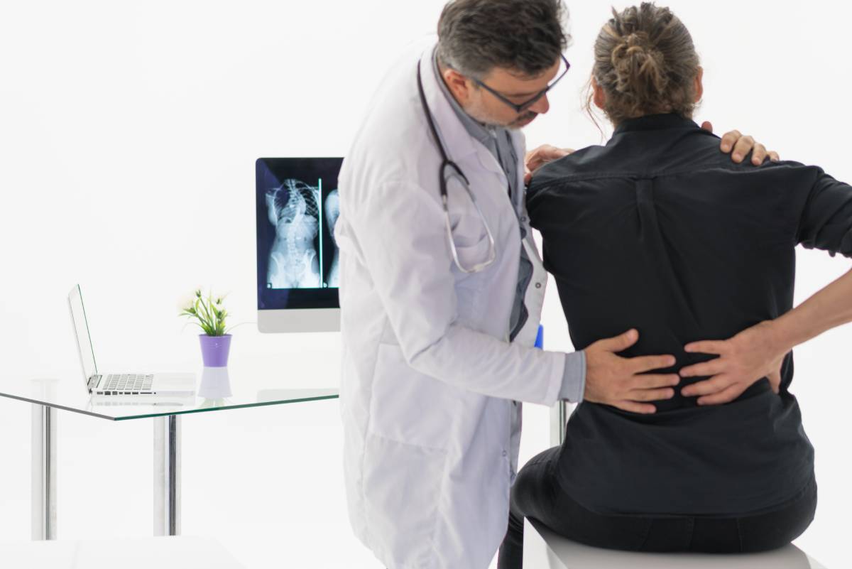
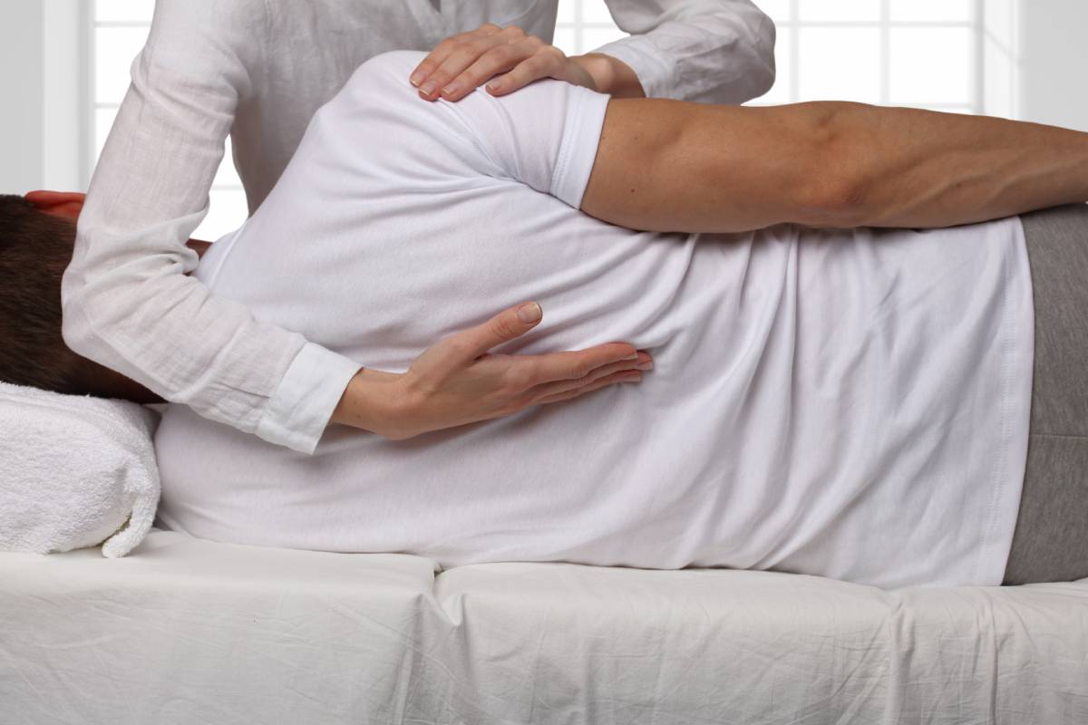
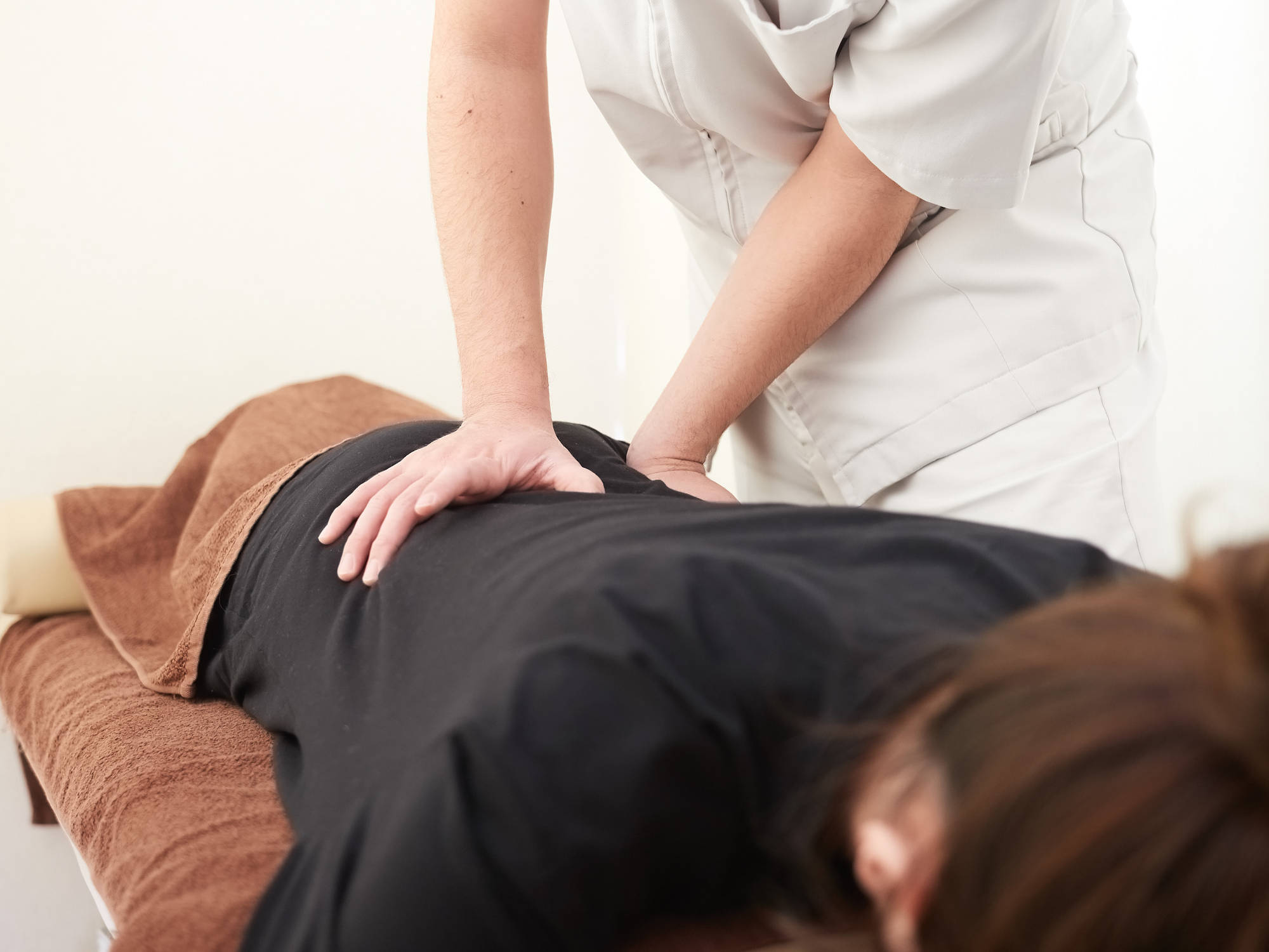
Migraine: Risk Factors and Management

The Global Burden of Diseases, Injuries, and Risk Factors (GBD) Study estimates that over one billion people had a migraine headache in 2016. This burden is significant, and migraines are ranked among the most common causes of “years of life lived with disability” (YLDs). Migraines caused 45.1 million YLDs globally in 2016, with the burden falling most heavily on women aged 15-49 years old. In this age group, migraines and tension-type headaches account for over ten percent of all YLDs.1 The management of migraine is also difficult, due to an incomplete understanding of their mechanism.

Researchers have achieved major advancements in understanding the pathophysiology of migraines, which are now recognized as manifestations of nervous system dysfunction rather than mere vascular headaches; while changes in blood flow to the brain perhaps contribute to pain, they likely do not initiate it. Diverse clinical features (including premonitory symptoms, aura, nausea, and dizziness) demonstrate the complexity of the condition. At least 38 genetic loci have been associated with migraines, but their functions differ; it is highly likely that complex gene-environment interactions cause migraine headaches and therefore must be considered in their management.2

Stress, exacerbating medications, metabolic changes associated with diet and neuroendocrine function, and hormonal changes (especially those induced by pregnancy and menstruation) may all also play a role in stimulating migraines.2 In some individuals, the attack frequency of episodic migraine can increase to the point of chronic migraine (at least 15 headache days per month for 3 months, with at least 8 of the headache days fitting the criteria for migraine headaches). Risk factors for chronification include overuse of acute migraine medication, obesity, depression, and stressful life events. Low socioeconomic and education statuses are associated with migraine chronification as well.3

Triptans and non-opioid analgesics (nonsteroidal anti-inflammatory drugs) are common medications used to treat migraines.2 However, the overuse of acute migraine medication (analgesic intake on more than 15 days per month or triptan intake on more than 10 days per month) is considered to be likely the most important cause of migraine progression, and therefore their usage must be carefully monitored.3

Beta-blockers are used as a preventative measure to limit the frequency of migraine. Local anesthetics (with or without steroids) are also sometimes injected in headache centers as preventative measures, particularly in the regions of the occipital nerves.2 Additional management options exist for those with chronic migraine: In 2010, the U.S. Food and Drug Administration approved Botox for the management of chronic migraine. The injection blocks the release of neurotransmitters implicated in perceptions of pain and has been shown to be an effective treatment, even for patients with concomitant medication overuse.4 Still, studies show that reducing medication overuse leads to significant migraine alleviation in chronic migraine patients.3

Because many people who experience migraines also report musculoskeletal issues like neck pain (reported by 75 percent of patients), there has been interest in incorporating chiropractic practices into neurological treatment plans. Harvard Medical School established the Osher Clinical Center (OCC) for Complementary and Integrative Therapies in 2007 at Brigham and Women’s Hospital, making it one of the first integrative medicine clinics at a tertiary care academic medical center. A 2019 case series published by Dr. Carolyn Bernstein and OCC researchers shared three cases demonstrating improvements in pain scores, increases in pain-free days, decreased medication usage, and decreased patient-reported anxiety/dysthymia with integrative approaches to treatment. The treatments included soft tissue therapies (including myofascial release, massage, and trigger point therapies), as well as spinal manipulation, which has been hypothesized to activate descending pain inhibitory pathways responsible for pain modulation.5

A 2019 systematic review that included six randomized clinical trials found that spinal manipulation may be an effective therapeutic treatment to reduce pain levels and slightly decrease migraine days. Larger-scale studies, however, are needed to further understand how chiropractic treatment may benefit those afflicted with migraine headaches.6

References

  1. GBD 2016 Headache Collaborators. Global, regional, and national burden of migraine and tension-type headache, 1990-2016: a systematic analysis for the Global Burden of Disease Study 2016. Lancet Neurol. 2018;17(11):954-976. https://thelancet.com/journals/laneur/article/PIIS1474-4422(18)30322-3/fulltext
  2. Charles A. The pathophysiology of migraine: implications for clinical management. Lancet Neurol. 2018;17(2):174-182.
  3. May A, Schulte LH. Chronic migraine: risk factors, mechanisms and treatment. Nat Rev Neurol. 2016;12(8):455-464.
  4. Escher CM, Paracka L, Dressler D, Kollewe K. Botulinum toxin in the management of chronic migraine: clinical evidence and experience. Ther Adv Neurol Disord. 2017;10(2):127-135.
  5. Bernstein C, Wayne PM, Rist PM, Osypiuk K, Hernandez A, Kowalski M. Integrating chiropractic care into the treatment of migraine headaches in a tertiary care hospital: A case series. Glob Adv Health Med. 2019;8:2164956119835778.
  6. Rist PM, Hernandez A, Bernstein C, et al. The impact of spinal manipulation on migraine pain and disability: A systematic review and meta-analysis. Headache. 2019;59(4):532-542.

Physiological Roles of Cholesterol

While much of the attention surrounding cholesterol relates to its negative association with heart disease, the molecule plays a number of essential physiological roles in promoting the health of humans and a diverse array of life forms — even fossils nearly 600 years old have been found to contain this lipid.1

Cholesterol, a waxy, light-colored fat, is an essential component in cell membranes. Flexible, porous plasma membranes surround every cell in the human body, and cholesterol helps maintain the membranes’ structure.2 Furthermore, the chemical properties of cholesterol allow a diverse array of signaling proteins to associate with the cell membrane, allowing cells to communicate with each other. Therefore, cholesterol has implications for reproductive biology (through sperm activation), cancer (through oncogenic protein regulation), and numerous other fields.3 Cholesterol is needed to create steroid-based hormones, including testosterone and progesterone. Other hormones like aldosterone, key for kidney functioning, and cortisol, which activates the body’s stress response, also require cholesterol for their manufacturing. So too does vitamin D, which is present in few foods but is critical for the body’s ability to use calcium. Cholesterol also plays a notable role in the formation of bile acids, which help the body digest and absorb fats.4

Thus, cholesterol, cholesterol metabolites, and precursors of cholesterol are critical for routine and essential bodily functions,3 including other, less commonly cited roles. Cells in the lens of the human eye contain enormously high levels of cholesterol, and this cholesterol is immensely important: those with genetic disorders affecting the cholesterol synthesis pathway often have cataracts, and cholesterol-lowering drugs have been associated with cataract-like effects in animals. Many studies have shown that statins that block cholesterol synthesis also are associated with cataracts.5

Unlike other parts of the body, the eye lens is avascular — not connected to the bloodstream — and high concentrations of cholesterol are not abnormal.5 Excess cholesterol in the bloodstream, however, can lead to atherosclerosis, or the formation of plaques in the walls of blood vessels. This can weaken the blood vessels, cause the formation of blood clots, and result in heart disease, stroke, or damage to the kidneys and intestines.4 Still, the impact of cholesterol is not fully explained by its quantity; the way it is transported matters, too. There are five major transporter proteins (called lipoproteins, which allow the lipid-soluble cholesterol to flow through the water-soluble bloodstream). While low-density lipoprotein (LDL) particles deliver cholesterol to tissues and are rich in pure cholesterol, high-density lipoprotein (HDL) particles help remove cholesterol from circulation, returning it to the liver.2

It is becoming increasingly clear that a full lipid profile test is critical for assessing cardiovascular disease (CVD) risk. If the proportion of HDL to LDL cholesterol is high, risks of atherosclerosis may be lower, even if someone has high total cholesterol.4 This is just one example of the complex considerations surrounding cholesterol management. The impact of cholesterol on bone health is another example of this. While studies in animals and cell cultures suggest that the cholesterol-lowering effects of some statins benefit bone metabolism, this relationship remains unclear in humans. It is widely known that cholesterol influences hematopoietic stem cell proliferation, driving the production of blood cells (a crucial life process) but this can also potentially contribute to the development of atherothrombosis (the accumulation of lipids and other materials that can form a clot in the arterial wall).6

Cholesterol performs many critical roles in the human body; still, its confounding detrimental health impact means that much focus has been placed on limiting its dietary intake.2 Historically, nutrition guidelines for reducing CVD risk have included explicit limitations for dietary cholesterol. However, the American Heart Association and American College of Cardiology have more recently eliminated specific targets because of the complexity of the relationship between CVD risk and cholesterol.7 Roughly 80 percent of cholesterol is created by the human body itself,2 a fact that serves as testament to its importance.

References 

  1. Davis J. Fat molecules preserved in a 558-million-year-old fossil help settle decades-old debate. National History Museum. Published 2018. https://www.nhm.ac.uk/discover/news/2018/september/fat-molecules-preserved-in-a-558-million-year-old-fossil-help-se.html  
  1. Corliss J. How it’s made: Cholesterol production in your body. Harvard Health Publishing. Published 2019. https://www.health.harvard.edu/heart-health/how-its-made-cholesterol-production-in-your-body  
  1. Tabas I. Cholesterol in health and disease. J Clin Invest. 2002;110(5):583-590. 
  1. Mulryan C. The role of cholesterol. Independent Nurse. Published 2012. https://www.independentnurse.co.uk/clinical-article/the-role-of-cholesterol/63612/  
  1. Widomska J, Subczynski WK. Why is very high cholesterol content beneficial for the eye lens but negative for other organs? Nutrients. 2019;11(5):1083. 
  1. Yin W, Li Z, Zhang W. Modulation of bone and marrow niche by cholesterol. Nutrients. 2019;11(6):1394. 
  1. Carson JAS, Lichtenstein AH, Anderson CAM, et al. Dietary cholesterol and cardiovascular risk: A science advisory from the American heart association. Circulation. 2020;141(3):e39-e53.  

The Link Between the Digestive and Nervous Systems

The link between the digestive and nervous systems, often termed the gut-brain axis (GBA) [1-4,6,7], consists of three parts—the enteric nervous system (ENS), “a collection of neurons in the gastrointestinal tract”; the central nervous system (CNS), comprising the brain and spinal cord; and the autonomous nervous system, which transfers information from the gut to the brain and vice versa [6]. These bidirectional interactions control “the motility, exocrine and endocrine secretions, and microcirculation of the gastrointestinal (GI) tract” [6], while also regulating the immune and inflammatory processes [4,5,6]. Researchers also posit that the GBA “supports and influences mood, cognitive function, and motivated behavior” [1].

One of the major indicators of gut health is its vast and complex microbial ecosystem, known as the gut microbiota [3]. Consisting mostly of bacteria, the gut microbiota plays a vital role in the bidirectional interactions that occur within the GBA [3]. For example, “it interacts with [the] CNS by regulating brain chemistry and influencing neuro-endocrine systems associated with stress response, anxiety, and memory function” [2]. Other functions include maintaining normal mucosal immune activity, “protecting against pathogens, participating in the intake of nutrients from the diet, metabolizing certain drugs and carcinogens, and influencing the absorption and distribution of fat” [3].

Studies indicate that disruption of the microbiota, known as dysbiosis, “perturbs host functions and, in some cases, causes the expression of overt and serious diseases such as IBD (inflammatory bowel disease) and Clostridium difficile colitis” [3]. According to Collins et al, “evidence is emerging of dysbiosis in patients with IBS (irritable bowel syndrome)” [3]. Dysbiosis has also been shown to “alter brain function and trigger the development of psychiatric disorders such as depression, schizophrenia, and Parkinson’s disease” [1].

Similarly, stress has been shown to produce adverse effects on the gut, and exacerbate the expression of certain diseases, such as IBD, and functional gastrointestinal disorders [7]. Additionally, “disorders of the enteric nervous system may result in motor, secretory, and inflammatory and immunologic dysfunction of the gut” [6]. However, some researchers postulate that “psychiatric and/or neurological diseases might be treated by using special probiotic bacteria,” special microbes (bacteria or yeast) which have been shown to improve the function of organs, the immune system, and the CNS [2,3].

Although the link between the digestive and nervous systems is directly related to overall health, “much remains to be elucidated with regard to the mechanism and impact of the interaction between the nervous system and the gastrointestinal system” [1]. More research is needed to determine the extent to which gut health influences the rest of the body and may be utilized to help treat certain conditions [1,6].

References 

1. Arneth, B. M. (2018). Gut-Brain Axis Biochemical Signaling from the Gastrointestinal Tract to the Central Nervous System: Gut Dysbiosis and Altered Brain Function. Postgraduate Medical Journal. DOI: 10.1136/postgradmedj-2017-135424 

2. Carabotti, M., Scirocco, A., et al. (2015). The Gut-Brain Axis: Interactions Between Enteric Microbiota, Central and Enteric Nervous Systems. Annals of Gastroenterology, 28(2), 203–209. PMID: 25830558. Available: link

3. Collins, S. M., & Bercik, P. (2009). The Relationship Between Intestinal Microbiota and the Central Nervous System in Normal Gastrointestinal Function and Disease. Gastroenterology, 136(6), 2003–2014. DOI: 10.1053/j.gastro.2009.01.075 

4. Costa, M., Brookes, S., et al. (2000). Anatomy and Physiology of the Enteric Nervous System. GUT, 47, 15–19. DOI: 10.1136/gut.47.suppl_4.iv15 

5. Furness, J. B. (2012). The Enteric Nervous System and Neuro-gastroenterology. Nature Reviews. Gastroenterology & Hepatology, 9(5), 286–294. DOI: 10.1038/nrgastro.2012.32 

6. Goyal, R. K., & Hirano, I. (1996). The Enteric Nervous System. New England Journal of Medicine, 334, 106–115. DOI: 10.1056/NEJM199604253341707 

7. Van Oudenhove, D. L. K., Tack, J., et al. (2004). Central Nervous System Involvement in Functional Gastrointestinal Disorders. Best Practice & Research Clinical Gastroenterology, 18(4), 663–680. DOI: 10.1016/j.bpg.2004.04.010 

Inflammation: Carbohydrates and Chiropractic Care

Inflammation contributes to many common chronic diseases, including obesity, type 2 diabetes, cardiovascular disease, arthritis, and various cancers [1, 2, 3]. Along with being implicated in the development of various diseases, inflammation also leads to an augmented risk of all-cause mortality during old age [2]. Consequently, the management of inflammation is of utmost concern to healthcare providers. This article will discuss two treatments that can reduce chronic inflammation: altered carbohydrate consumption and chiropractic care.

Carbohydrate consumption can be measured by two markers. Glycemic index (GI) quantifies the propensity for an individual’s carbohydrate intake to increase one’s blood glucose level [1] Glycemic load (GL) is the product of an individual’s total carbohydrate intake and dietary GI [1]. Consistent consumption of high-GI foods often leads to acute and chronic inflammation [2]. Additionally, several studies document the positive correlation between dietary measures of GI/GL and levels of high-sensitivity C-reactive protein (hsCRP), a marker of inflammation [2]. Therefore, one way to reduce inflammation is by avoiding high-GI/GL foods, such as breakfast cereals, potatoes, and sweet treats [4].

Another way to lower inflammation is by consuming fewer refined carbohydrates [2, 3]. Refined carbohydrates not only promote postprandial inflammation, but also activate neural addiction pathways, reduce energy expenditure, and contribute to weight gain [3]. Individuals should replace refined carbohydrates with whole-grain foods. Whole-grain foods are rich in bioactive compounds that combat inflammation through antioxidant enzyme activation and free radical scavenging [2]. While low-GI/GL diets are associated with anti-inflammatory benefits much more consistently, whole-grain diets are still strongly tied to reduced inflammation [2].

Chiropractic care can also reduce inflammation. Chiropractors may advise their clients to make dietary changes in line with the recommendations above, but they can also reduce inflammation by engaging in specific spinal manipulations [5]. When some experiments began reporting how chiropractic treatments reduced the painful effects of inflammation, the connection between the two became clear. One such study, conducted by Song et al. in 2006, measured the impact of activator-assisted spinal manipulative therapy (ASMT) on rats suffering from acute intervertebral foramen inflammation. When ASMT was applied on L5 and/or L6 spinous processes, the rats experienced shorter and less severe thermal and mechanical hyperalgesia [6].

Various experiments indicate that chiropractic treatments can directly reduce inflammation. A blinded study consisting of 21 patients sought to compare the effect of chiropractic spinal manipulations on two inflammation markers: CRP and interleukin-6 (IL-6) [5]. Two weeks after receiving nine spinal manipulations, patients exhibited significantly reversed inflammatory processes compared to the control group [5]. A larger experiment documented how a single session of spinal manipulation therapy reduced subjects’ levels of two inflammatory cytokines, tumor necrosis factor α and interleukin 1β [7]. In both cases, chiropractic spinal manipulations proved to be a promising treatment against inflammation.

Despite the complex and confounding molecular interactions associated with inflammation, researchers have identified certain dietary and chiropractic adjustments that can successfully reduce low-grade inflammation.

References 

[1] L. Galland, “Diet and Inflammation,” Nutrition in Clinical Practice, vol. 25, no. 6, p. 634-640, Dec 2010. [Online]. Available: https://doi.org/10.1177/0884533610385703.  

[2] A. E. Buyken et al., “Association between carbohydrate quality and inflammatory markers: systematic review of observational and interventional studies,” American Journal of Clinical Nutrition, vol. 99, no. 4, p. 813-833, Apr 2014. [Online]. Available: https://doi.org/10.3945/ajcn.113.074252.  

[3] D. Seaman, “Weight gain as a consequence of living a modern lifestyle: a discussion of barriers to effective weight control and how to overcome them,” Journal of Chiropractic Humanities, vol. 20, no. 1, p. 27-35, December 2013. [Online]. Available: https://doi.org/10.1016/j.echu.2013.08.001.  

[4] L. Richards, “What are high and low glycemic index foods?,” Medical News Today, Updated February 7, 2021. [Online]. Available: https://www.medicalnewstoday.com/articles/high-glycemic-index-foods.  

[5] R. A. Roy, J. P. Boucher, and A. S. Comtois, “Inflammatory response following a short-term course of chiropractic treatment in subjects with and without chronic back pain,” Journal of Chiropractic Medicine, vol. 9, no. 3, p. 107-114, September 2010. [Online]. Available: https://doi.org/10.1016/j.jcm.2010.06.002.  

[6] X. J. Song et al., “Spinal Manipulation Reduces Pain and Hyperalgesia After Lumbar Intervertebral Foramen Inflammation in the Rat,” Journal of Manipulative and Physiological Therapeutics, vol. 29, no. 1, p. 5-13, January 2006. [Online]. Available: https://doi.org/10.1016/j.jmpt.2005.10.001.  

[7] J. A. Teodorczyk-Injeyan, J. S. Injeyan, and R. Ruegg, “Spinal Manipulative Therapy Reduces Inflammatory Cytokines but Not Substance P Production in Normal Subjects,” International Conference on Chiropractic Research, vol. 29, no. 1, p. 14-21, January 2006. [Online]. Available: https://doi.org/10.1016/j.jmpt.2005.10.002.  

Flexion Distraction Manipulation for Disk Pain

The human spine is composed of bones (vertebrae) that are stacked on top of each other and cushioned by rubbery pads that lie between them, called “spinal disks” or intervertebral disks. The spinal disks are roughly a quarter of an inch thick, with an elastic, fluid-filled core. They help prevent bone-on-bone friction and also increase the spine’s shock-absorption and flexibility. However, when subject to stress, the material inside the spinal disks can swell, bulging out from between the vertebrae and pressing against the surrounding nerves.1 This “herniated disk” is a common cause of lower back pain, and flexion distraction manipulation is one way to treat it.2

Flexion distraction manipulation is a chiropractic therapy in which the patient lies face-down on a mobile table, which gently stretches the spine while the practitioner massages key areas of the back. (“Flexion” refers to bending the body in such a way that brings joints closer together, and “distraction” refers to the pressure put on the body.) This creates a negative pressure that pulls the disk back between the vertebrae. It also prevents the tough, circular exterior of the spinal disk, the annulus fibrosus, from becoming distorted, which can cause pain. Since its invention in the 1960s by James Cox, flexion distraction has been the subject of various case studies and anecdotal reports, which have helped characterize and clarify its therapeutic benefits.2

For example, a 2019 study published in the Journal of Physical Therapy Science by Oh et al. examined the clinical outcomes of 30 female patients who visited a South Korean orthopedic clinic with symptoms consistent with herniated intervertebral disks. The researchers administered flexion distraction and another, similar chiropractic therapy (the drop technique) to two groups of 15 herniated disk patients. The control group received spinal decompression — non-chiropractic therapy that uses a specialized medical device instead of manual therapy. The researchers found that the patients’ straight leg raising angle and intervertebral disk height significantly improved in both groups, but that there was no significant difference in the increase when comparing the groups — in other words, the treatments were equally effective.3

The effectiveness of flexion distraction for patients with herniated disks has been repeatedly demonstrated. Kwon et al., cited by Oh, also found improved straight leg raising angles after application of flexion distraction.3 A study by Gudavalli et al. of cadaveric spines found that spinal disk space increased by approximately 3 mm and the angle of the intervertebral disk by around 6 degrees in the lumbar vertebrae of cadavers.4 In the study by Oh et al., the disk space increased by an average of 1.3 mm.3 Gay et al. found in another cadaveric study that the technique decreased the pressure within the intervertebral disk by 65 percent.5

Ultimately, patients may choose flexion distraction manipulation over another type of therapy based on their specific ailment. Choi et al. examined the effects of flexion distraction manipulation in 30 patients with lumbar spinal stenosis (narrowing of the spinal canal, which can be due to disk bulging), finding that while both pain and disability decreased in both groups, the decreases were more significant in the flexion distraction group in an intergroup comparison.6 This could support the use of flexion distraction for spinal stenosis over other therapies — spinal decompression therapy, for example, is not recommended for patients with spinal stenosis. Similarly, spinal decompression therapy is not recommended for spondylolisthesis and ankylosing spondylitis, but flexion distraction may be advisable.7

While flexion distraction manipulation remains a popular treatment option for a variety of conditions, including those involving disk pain, scientific studies evaluating its efficacy remain limited. A 2005 literature review of flexion distraction manipulation identified 30 articles on the topic, most of which were case reports or studies with small sample sizes. The article concluded that further investigation is needed “to establish the efficacy and safety of distraction manipulation and to explore biomechanical, neurological, and biochemical events that may be altered by this treatment.”8

References

1. Wheeler T. Understanding basic information about spinal disk problems. Webmd.com. Published 2019. https://www.webmd.com/pain-management/understanding-spinal-disk-problems-basic-information

2. Merckling J. Flexion distraction to the rescue for chronic back pain. Mercklingdc.com. Published 2018. https://mercklingdc.com/2018/10/29/flexion-distraction-to-the-rescue-for-chronic-back-pain/

3. Oh H, Choi S, Lee S, Choi J, Lee K. Effects of the flexion-distraction technique and drop technique on straight leg raising angle and intervertebral disc height of patients with an intervertebral disc herniation. J Phys Ther Sci. 2019;31(8):666-669.

4. Gudavalli MR, Cambron JA, McGregor M, et al. A randomized clinical trial and subgroup analysis to compare flexion-distraction with active exercise for chronic low back pain. Eur Spine J. 2006;15(7):1070-1082.

5. Gay RE, Ilharreborde B, Zhao KD, Berglund LJ, Bronfort G, An K-N. Stress in lumbar intervertebral discs during distraction: a cadaveric study. Spine J. 2008;8(6):982-990.

6. Choi J, Lee S, Jeon C. Effects of flexion-distraction manipulation therapy on pain and disability in patients with lumbar spinal stenosis. J Phys Ther Sci. 2015;27(6):1937-1939.

7. Spinal decompression vs chiropractic flexion-distraction technique. Evergreenclinic.ca. Published September 26, 2020. https://evergreenclinic.ca/difference-between-chiropractic-flexion-disctraction-technique-and-spinal-decompression/

Gay RE, Bronfort G, Evans RL. Distraction manipulation of the lumbar spine: a review of the literature. J Manipulative Physiol Ther. 2005;28(4):266-273.

Use of Gait Analysis to Assess Biomechanical Abnormalities

Walking is a motion that requires accurate coordination of a number of different musculoskeletal groups. For this reason, abnormalities in a patient’s gait can be highly informative of a number of different issues, from diabetic foot complications to acute sports injuries. Gait analysis is a practice that was first introduced in the 1970s and has since been used to clinically investigate the source of nervous, skeletal, or muscular afflictions, as well as biomechanical abnormalities in the feet, ankles, legs, knees, hips, and back. It is used as both a preventative tool and a measurement of progress for patients undergoing treatment. 

Gait analysis can be performed using a wide range of technology, and methodology has continually improved throughout recent years. i,ii Footstep analysis can be performed using a pressure-sensitive compression pad, which is used to quantify stride length and stepping pace while also capturing impressions of a patient’s footfall. Such analysis can be highly informative of an individual’s balance and the overall consistency of their gait. iii Force and pressure measurements may be taken using a pressure-sensitive walkway attached to a computer, which then generates images to visualize a patient’s biomechanics. Scanning electronics and 3D sensors have also been used to help measure asymmetrical gait patterns. However, the most common and cost-effective form of gait analysis involves taking video footage of the patient walking in various directions and from different angles. iii The patient may walk on a flat surface or a treadmill, where incline and speed can be manually manipulated, and external motion sensors may also be worn for quantitative measurements. 

Specific trends in gait abnormalities have been associated with a number of different conditions. For example, one study found that patients afflicted with spinal disorders had significantly slower gait speed and step length, as well as increased step width, as compared to controls.i Another study established specific kinematic and kinetic gait parameters for patients with cervical spondylotic myelopathy, in which the authors noted significant decreases in knee flexion during swing, total sagittal knee range of motion, and peak ankle plantar flexion.ii Raccagni et al. found that gait analysis could successfully distinguish between idiopathic Parkinson’s disease and atypical parkinsonian disorders, suggesting a high degree of diagnostic accuracy.iii  

These findings demonstrate that gait analysis provides a highly nuanced way of characterized biomechanical abnormalities and can be used in both a diagnostic manner and as a measure of disease or recovery progression. It is therefore of great clinical use to perform gait analysis on patients with lower-body and spinal afflictions alongside other diagnostic tests.  

References 

i. Haddas, R., Ju, K.L., Belanger, T. et al. The use of gait analysis in the assessment of patients afflicted with spinal disorders. European Spine Journal 27, 1712–1723 (2018). https://doi.org/10.1007/s00586-018-5569-1 

ii. Malone, A., Meldrum, D., & Bolger, C. (2012). Gait impairment in cervical spondylotic myelopathy: comparison with age- and gender-matched healthy controls. European Spine Journal 21(12), 2456–2466. https://doi.org/10.1007/s00586-012-2433-6 

iii. Raccagni, C., Gaßner, H., Eschlboeck, S., Boesch, S., Krismer, F., Seppi, K., Poewe, W., Eskofier, B. M., Winkler, J., Wenning, G., & Klucken, J. (2018). Sensor-based gait analysis in atypical parkinsonian disorders. Brain and Behavior 8(6), e00977. https://doi.org/10.1002/brb3.977 

Vertigo Management with Chiropractic Care

Vertigo is an abnormal sensation of motion or loss of balance and can be either chronic or intermittent. Brief sensations of vertigo may arise for a wide variety of reasons, such as sitting up quickly or playing three-dimensional video games. More persistent forms of vertigo usually arise due to dysfunction of the inner ear, which is responsible for our sense of balance and positioning. Cold viruses, head trauma, and Meniere’s disease are all conditions that affect the inner ear and can cause a sensation of unsteadiness (The University of Iowa 2018). Musculoskeletal conditions, including nerve damage to the legs, muscle weakness, and joint instability, may also give rise to vertigo and cause difficulties with movement. Finally, certain conditions such as Parkinson’s disease, depression, and impaired vision can be catalysts for vertigo (Mayo Foundation for Medical Education and Research 2020). 

It is extremely rare for young children to be affected by vertigo, and it almost always occurs in adulthood. Approximately one in 20 adults experience vertigo each year, and the majority of people with the condition find that it severely impairs their daily activities and ability to work (van Vugt 2017). Furthermore, falls are the leading cause of injury among senior citizens and can cause severe trauma such as fractures. As falls are largely caused by impairments in balance, the potential consequences of vertigo become more severe with age.  

If vertigo is accompanied by tinnitus, ear pressure, or hearing loss, the inner ear may be the source of the condition. An otolaryngologist, a specialist in ear disorders, may examine the onset, duration, and severity of ear discomfort experienced. Other symptoms that commonly accompany ear disorders include nausea and vomiting, as well as lowered heart rate (The University of Iowa 2018). Positional vertigo, a form of vertigo in which symptoms change depending on the position of the head, is common and primarily originates in the inner ear. 

Vertigo is usually treatable with physical therapy, medication, and/or surgery (The University of Iowa 2018). Neurologists typically order tests and scans to determine the cause of the condition before developing a treatment plan. Symptoms of vertigo caused by more serious structural problems may require surgery, while in some cases, balance exercises and lifestyle changes alone can help manage symptoms. Other conservative treatments, such as restricting foods and drinks that cause migraines or impair the senses (e.g. alcohol and coffee), may be recommended by a medical professional. 

Chiropractic care is another effective form of treatment for vertigo that uses hands-on manipulation to help patients improve their balance and coordination. According to a 2010 survey conducted by the National Board of Chiropractic Examiners, chiropractors report seeing, on average, between one and three patients per month for concerns about dizziness (Ndetan 2016). Some studies suggest that chiropractic manipulations targeting the cervical spine may be helpful in treating balance disorders, such as vertigo, by re-positioning the neck in its optimal location and bringing the body back to equilibrium.  

A 2009 study examined the effects of spinal manipulation and manual therapy on dizziness and balance at a chiropractic college health center and a senior fitness center (Strunk 2009). A group of 19 adults, aged 40 years or older with a median age of 70, completed the study. All patients were treated by either a clinician or a chiropractic student intern twice per week, each session lasting 15-20 minutes, during an 8-week intervention period. The Dizziness Handicap Inventory, the Short Form Berg Balance Scale (SF-BBS), and the Neck Disability Index were used to measure the effects of the treatments. A large difference in the SF-BBS before and after the intervention period was measured in most patients, demonstrating an improvement in balance. Some patients also showed reduced dizziness and neck pain at the conclusion of the study. 

References 

Mayo Foundation for Medical Education and Research. (2020). Balance Problems. Mayo Clinic. https://www.mayoclinic.org/diseases-conditions/balance-problems/symptoms-causes/syc-20350474#

Ndetan, H., et al. (2016). The Role of Chiropractic Care in the Treatment of Dizziness or Balance Disorders: Analysis of National Health Interview Survey Data. J Evid Based Complement Altern Med, 21:138–142. doi:10.1177/2156587215604974.  

Strunk, R., et al. (2009). Effects of Chiropractic Care on Dizziness, Neck Pain, and Balance: A Single-Group, Preexperimental, Feasibility Study. Journal of Chiropractic Medicine, 8(4), 156–164. doi:10.1016/j.jcm.2009.08.002.  

The University of Iowa. (2018). Vertigo: Frequently Asked Questions. The University of Iowa Hospitals & Clinics. https://uihc.org/health-topics/vertigo-frequently-asked-questions.  

van Vugt, V., et al. (2017). Chronic Vertigo: Treat with Exercise, Not Drugs. BMJ Publishing Group, 358. doi:10.1136/bmj.j3727. 

Nerve Compression: Symptoms and Management

Nerve compression, or nerve entrapment, “is a condition caused by direct pressure on a nerve” [5], often occurring in the wrists, elbows, feet, or spine. Symptoms may include “pain, weakness, or paresthesia (“pins and needles”) [5], ranging from mild to severe, and can develop suddenly or gradually [7]. Patients suffering from nerve compression may also experience a dull, aching sensation radiating from the origin of the compressed nerve, while others may experience numbness [1,3,7]. For patients presenting with these symptoms, especially in the “absence of a known bone, soft tissue, or vascular injury,” researchers have suggested that nerve entrapment syndromes be considered [5].

Nerve entrapment syndromes tend to affect those whose occupations involve repetitive movements or the lifting of heavy objects, such as athletes, office workers, and musicians [1]. Common nerve entrapment syndromes include carpal tunnel syndrome, radial nerve entrapment, ulnar entrapment, and spinal cord compression.

Carpal tunnel syndrome is produced by compression of the median nerve at the wrist [1]. Affected patients report numbness, tingling, and pain in the hand, which often worsens at night or after use of the hand [1]. Similarly, patients with ulnar nerve entrapment or radial nerve entrapment, resulting from compression of the ulnar and radial nerves, respectively, generally describe numbness or pain along the forearms, hands, and fingers [1,3,5]. Symptoms of spinal nerve compression, caused by pressure on the spinal cord, typically resulting from general wear and tear or osteoporosis, may consist of pain and stiffness in the back or neck; “numbness, cramping, or weakness in the arms, hands, or legs; intense pain spreading to the arms, buttocks, or legs (sciatica); and loss of sensation in the feet” [7].

Surgical methods for treating nerve compression, such as decompressions [2,4,6] and the sectioning of ligaments [1], have shown strong success rates and, as advised by Jacobsen et al., “should be considered in patients with persistent pain and identifiable sources of entrapment” [4]. However, while surgical treatments for nerve compression typically yield positive results, some experts consider it to be a last resort, as nonsurgical treatments have proven to be effective in many situations [4,7].

Non-surgical treatments for managing nerve compression may involve splints, pharmacological interventions (NSAIDs), ultrasound therapy, steroid injections, physical therapy, and rest [1]. A study on entrapment neuropathies in the upper extremities recommends rest, splints, and anti-inflammatories, specifically for patients with carpal tunnel syndrome who experience “acute flare-ups, and in those with minimal to intermittent symptoms” [1]. Another study, citing research on the efficacy of splinting when combined with local steroid injections, reports that “22 percent [of patients] were free of symptoms at the end of a year-long trial.”

Chiropractic techniques have also been used to help manage nerve compression. A 2008 study noted a patient with carpal tunnel syndrome who, upon receiving chiropractic manipulation therapy in combination with other techniques over a period of nine months, was able to “return to occupational and social activities” [6]. The research performed by Jefferson-Falardeau et al., focusing on chiropractic management of a patient with radial nerve entrapment syndrome, found that the patient benefited from chiropractic management using standard chiropractic, applied kinesiology, and neural mobilization techniques” [5]. Similarly, the study performed by Illes et al. concluded that “chiropractic treatment consisting of manipulation, soft tissue mobilizations, exercise, and education of workstation ergonomics appeared to reduce the symptoms of ulnar nerve compression symptoms” [3]. Chiropractic techniques have also been said to help treat spinal cord compression.

Treatments for managing nerve compression may be prescribed individually. However, research suggests that a multimodal therapeutic approach may improve results [6].

References

  1. Dawson, D. M. (1993). Entrapment Neuropathies of the Upper Extremities. New England Journal of Medicine, 329, 2013–2018. DOI: 10.1056/NEJM199312303292707
  2. Dhinsa, B. S., Hussain, L., et al. (2018). The Management of Dorsal Peroneal Nerve Compression in the Midfoot. The Foot, 35, 1–4. DOI: 10.1016/j.foot.2017.12.005
  3. Illes, J. D., & Johnson, T. L. Chiropractic Management of a Patient With Ulnar Nerve Compression Symptoms: A Case Report. Journal of Chiropractic Medicine, 12(2), 66–73. DOI: 10.1016/j.jcm.2013.03.002
  4. Jacobson, L., Dengler, J., et al. (2020). Nerve Entrapments. Clinics in Plastic Surgery, 47(2), 267–278. DOI: 10.1016/j.cps.2019.12.006
  5. Jefferson-Falardeau, J., & Houle, S. Chiropractic Management of a Patient With Radial Nerve Entrapment Symptoms: A Case Study. Journal of Chiropractic Medicine, 18(4), 327–334. DOI: 10.1016/j.jcm.2019.07.003
  6. McHardy, A., Hoskins, W., et al. (2008). Chiropractic Treatment of Upper Extremity Conditions: A Systematic Review. Journal of Manipulative & Physiological Therapeutics, 31(2), 146–159. DOI: 10.1016/j.jmpt.2007.12.004
  7. Spinal Cord Compressions. Johns Hopkins Medicine. https://www.hopkinsmedicine.org/health/conditions-and-diseases/spinal-cord-compression

Knee Pain Due to Pelvic Imbalance

Pelvic imbalance, also referred to as “pelvic tilt”, is a common condition affecting both men and women. Contributing factors include discrepancy between leg length, limited flexibility, lack of strength or muscular endurance, irregular posture, poor sitting habits, congenital scoliosis, flat feet, and improper footwear (Karp Rehabilitation 2018). Pelvic imbalance can be a minor issue and is even quite normal; many people have slight leg length discrepancies due to the random asymmetry of bilateral traits resulting naturally from the way an embryo develops. If, however, the degree of pelvic imbalance is too large, the body will try to correct itself by putting more pressure on certain areas; consequently, the strain placed on the back, stomach, pelvis region, and lower extremities can vary significantly from one side to the other. Pelvic imbalance may lead to severe issues such as joint misalignment, muscular imbalances, instability, abnormal posture, and pain in various parts of the body, including the knee (Karp Rehabilitation 2018). 

While pelvic misalignment is common in men, women are particularly vulnerable due to their larger and more mobile pelvises. A report funded by the National Institutes of Health found that nearly 24 percent of women in the U.S. are affected by one or more pelvic floor disorders and that the frequency of pelvic floor disorders increases with age (U.S. Department of Health and Human Services 2015). The imbalance is especially prevalent during pregnancy as a woman’s body releases hormones that relax ligaments to create space for the developing fetus. The increased width of the pelvis often destabilizes the spine and, due to the added pressure from the weight of the child, shifts the spine out of alignment. A 2019 study in which pelvic measurements were obtained from 201 women at 12, 24, 30, and 36 weeks of pregnancy, and 1 month after childbirth found significant differences in the measurements over time (Morino 2019). The anterior and posterior width of the pelvis—the distance between the bilateral anterior/posterior superior iliac spines—became significantly wider as pregnancy progressed. Furthermore, the degree of pelvic tilt of the anterior pelvis increased during pregnancy. The anterior width of the pelvis was not recovered even at one month post-childbirth. 

Knee pain is a common side effect of pelvic imbalance. A 2010 study examined the relationship between pelvis malposition and knee pain in 100 endurance runners, 50 with knee pain and 50 without knee pain, and concluded that there is a correlation between one-sided pelvic mispositioning and knee pain during activities such as endurance running (Siegele 2010). 

Understanding the causes of pelvic misalignment and devising a treatment plan may help avert knee complications or alleviate pain. One case report describes successful chiropractic treatment of a female patient’s knee pain stemming from pelvic imbalance (Bucek 2019). The patient complained of ongoing anteromedial knee pain when walking and a persistent feeling of her knee “giving out” after walking no more than a half mile. The patient was administered five treatments over the course of ten days, and in each treatment received chiropractic manipulation of the sacroiliac joint, kinesiology taping, and gluteus medius exercises to improve muscle strength. Following the treatments, she reported a resolution of her knee issues and experienced no pain while walking or performing other activities (Bucek 2019). 

As described, conservative measures to correct imbalance and resolve knee pain may be effective for patients experiencing knee complications stemming from pelvic misalignment. Chiropractic treatments may help increase muscle strength and flexibility as well as ease the tension on supportive tissues that are strained from misaligned or stiff pelvic joints. 

References 

Bucek, D. (2019). Reduction of Knee Pain in a 45-Year-Old Woman After Pelvic Manipulation and Kinesiology Taping: A Case Report. Journal of Chiropractic Medicine, 18(3), 236-241. https://doi.org/10.1016/j.jcm.2019.07.006. 

First Digital Solutions. (2016, October 14). Pelvic Imbalance. Chelsea Osteopathic Clinic. https://chelseaosteopaths.co.uk/pelvic-imbalance/.  

Karp Rehabilitation. (2018, February 20). Understanding Pelvic Injuries & How to Treat Them. Karp Rehabilitation. http://karprehab.com/pelvic-injuries/.  

Morino, S., et al. (2019). Pelvic alignment changes during the perinatal period. PloS one, 14(10), e0223776. https://doi.org/10.1371/journal.pone.0223776. 

Siegele, J., et al. (2010). Relation between pelvis malposition and functional knee pain by long distance running. Sportverletzung Sportschaden, 24(3), 144–149. https://doi.org/10.1055/s-0029-1245638. 

U.S. Department of Health and Human Services. (2015, September 28). Roughly One Quarter of U.S. Women Affected by Pelvic Floor Disorders. National Institutes of Health. https://www.nih.gov/news-events/news-releases/roughly-one-quarter-us-women-affected-pelvic-floor-disorders.  

The Effect of Spinal Manipulation on Shoulder Range of Motion

Shoulder pain is a common reason for people to seek medical care and is the third most common site of musculoskeletal pain, after the lower back and knee (1,2). In the United States alone, the direct cost of treatment for shoulder dysfunction in 2000 was estimated to be $7 billion. 1-2% of the population is estimated to experience shoulder pain each year. Beyond direct costs, regional pain in the shoulder can also evolve into more generalized pain syndromes, and pain can lead to limitations in range of motion (ROM), subsequently impairing functional activities (1,2). As a result, it is necessary to investigate whether existing treatments for improving shoulder range of motion and pain, such as spinal manipulation, are effective. 

Chiropractic care frequently draws on the theory of regional interdependence, which posits that deficiencies in one area of the body may be associated with primary symptoms in another area, despite being apparently unrelated (2). Previous studies found a decrease in shoulder pain immediately after thoracic manipulation but did not find changes in scapular kinematics or muscle activity (3). Due to the importance of shoulder ROM to daily life, a study by Silva et al. sought to investigate the effect of thoracic spinal manipulation (SM) on shoulder range of motion in people with chronic shoulder pain, with a secondary goal of decreasing pain levels (2). 

The study examined 60 participants who presented with shoulder pain for at least 6 months and who showed clinical signs of rotator cuff tendinopathy, through a combination of positive Hawkins, Neer, or Jobe tests; or pain/weakness with external rotation of the arm. The study excluded patients with contraindications to SM (such as a history of cancer, osteoporosis, or fracture), a pacemaker, or clinical signs of complete rotator cuff tear. On average, participants had experienced nearly four years of pain before entering the study (2). 

Participants were assigned to either the manipulation group or placebo group but were blinded to which group they belonged to. The manipulation group received two high-velocity low-amplitude thrusts between the T4 and T5 vertebrae, while the placebo group received a constant, low pressure contact while assuming the same prone position. Researchers evaluated three key metrics before and after the intervention: the degree of shoulder flexion, the degree of shoulder abduction, and self-reported shoulder pain (2). 

After thoracic spinal manipulation, participants demonstrated a statistically significant increase in both flexion and abduction range of motion in the painful shoulder, as well as a significant increase in abduction of the nonpainful shoulder. The improvement in abduction in the painful shoulder was also above the calculated minimal detectable change, giving this effect clinical significance as well. However, the placebo group also demonstrated a statistically significant improvement in shoulder ROM after receiving the placebo intervention. Both groups reported less pain after their respective interventions (p < 0.01), but this change was not clinically significant (2). 

This study demonstrated that thoracic spinal manipulation clinically improved abduction, one aspect of range of motion, in the painful shoulder, but that thoracic SM did not improve flexion or pain levels (2). Future research should include a true control group in order to test for a placebo effect, which is a potential explanation for changes experienced by the placebo group, as well as investigate other treatment techniques for shoulder pain. 

References 

1. Silva ACD, Santos GM, Marques CMG, Marques JLB. Immediate Effects of Spinal Manipulation on Shoulder Motion Range and Pain in Individuals With Shoulder Pain: A Randomized Trial. J Chiropr Med. 2019;18(1):19-26. doi:10.1016/j.jcm.2018.10.001 

2. Tekavec, E., Jöud, A., Rittner, R. et al. Population-based consultation patterns in patients with shoulder pain diagnoses. BMC Musculoskelet Disord. 2012;13(238). doi:10.1186/1471-2474-13-238 

3. Haik MN, Alburquerque-Sendín F, Silva CZ, et al. Scapular Kinematics Pre- and Post-Thoracic Thrust Manipulation in Individuals With and Without Shoulder Impingement Symptoms: A Randomized Controlled Study. J Orthop Sports Phys Ther. 2014;44(7):475-487. doi:10.2519/jospt.2014.4760