The One-To-Zero Technique in Individuals With Occipito-Atlantal Joint Dysfunction

The One-To-Zero Technique in Individuals With Occipito-Atlantal Joint Dysfunction

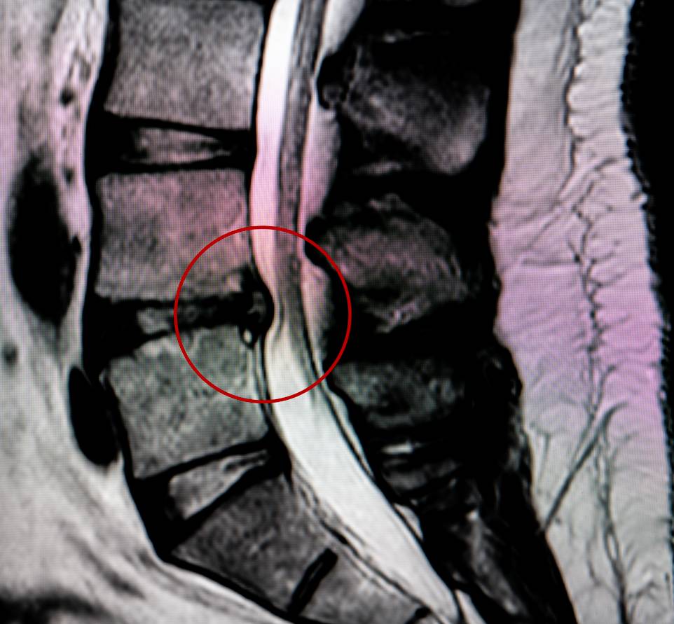
The “One-to-Zero Technique” is a unique method used primarily in chiropractic and osteopathic practices, focusing on treating occipito-atlantal joint dysfunction. This joint, located at the junction between the occipital bone of the skull and the first cervical vertebra, the atlas, is vital for the movement and stability of the head and neck. Dysfunction in this area can arise from various causes such as traumatic injuries, poor posture, congenital anomalies, or degenerative changes, leading to symptoms like neck pain, headaches, dizziness, and restricted head and neck movement.

The essence of the One-to-Zero Technique lies in its gentle approach to realigning the occipito-atlantal joint, aiming to move from a state of dysfunction (‘one’) to normal function (‘zero’). The process involves an initial assessment of the joint’s range of motion and pain level, followed by positioning the patient to relax the neck muscles for optimal access to the joint. The practitioner then uses specific hand placements and gentle pressure to correct misalignments, followed by a reassessment to evaluate changes in mobility and symptom relief.

While there is anecdotal evidence supporting the efficacy of this technique, comprehensive clinical research is somewhat limited. However, when performed by a qualified professional, it is generally considered safe, although it requires precise knowledge of neck anatomy due to the proximity to critical structures like arteries and nerves. The technique isn’t suitable for all cases of occipito-atlantal dysfunction; suitability depends on the specific cause and severity of the dysfunction.

Often, this technique is part of a broader treatment plan that includes physical therapy exercises to strengthen neck muscles, along with ergonomic adjustments and posture correction to prevent recurrence. Regular follow-ups with the practitioner are crucial for ensuring long-term effectiveness and addressing any recurring issues.

Despite its potential benefits, some in conventional medicine remain skeptical about the effectiveness of chiropractic and osteopathic manipulations, advocating for more evidence-based approaches. On the other hand, proponents of the technique emphasize a holistic approach to patient health, considering physical, emotional, and lifestyle factors. The One-to-Zero Technique represents a hands-on approach to addressing a condition that can significantly impact quality of life. While promising, it underscores the need for a tailored approach to each patient, grounded in a comprehensive understanding of their unique anatomical and health contexts. Future research and interdisciplinary collaboration could further clarify its place in the spectrum of healthcare options.

Lateral Epicondyle Tendinopathy

Lateral Epicondyle Tendinopathy

Chiropractic techniques have evolved to address a variety of musculoskeletal conditions, including lateral epicondyle tendinopathy, commonly known as tennis elbow. This condition is characterized by pain and tenderness on the outside of the elbow, often caused by overuse of the forearm muscles and tendons. The focus of chiropractic care in treating lateral epicondyle tendinopathy lies in alleviating pain, reducing inflammation, and promoting healing while improving joint function.

The initial approach in chiropractic treatment for lateral epicondyle tendinopathy typically involves a thorough assessment of the patient’s elbow joint, as well as the surrounding muscles and tendons. This assessment is crucial to identify the specific areas of dysfunction and to tailor a treatment plan that addresses the individual needs of the patient. Following the assessment, a chiropractor may employ a variety of hands-on techniques aimed at reducing tension and pain in the affected area.

One of the primary chiropractic techniques used is the adjustment or manipulation of the elbow joint. This technique involves applying a controlled, sudden force to the joint, aiming to improve range of motion and reduce pain. The adjustment can help to release joint restrictions and improve alignment, which in turn can alleviate stress on the tendons and muscles around the elbow.

Soft tissue therapies are also commonly used in treating lateral epicondyle tendinopathy. These may include massage therapy, which helps to relax tight muscles and improve blood flow to the area, aiding in the healing process. Myofascial release techniques can also be employed to release tension in the fascia – the connective tissue surrounding muscles – thereby reducing pain and improving mobility.

Another important aspect of chiropractic care for this condition is the use of modalities such as ultrasound therapy or low-level laser therapy. Ultrasound therapy uses sound waves to produce a deep heating effect that can reduce inflammation, promote blood flow, and speed up the healing process. Low-level laser therapy, on the other hand, uses light to enhance cellular function and promote tissue repair without generating heat. These modalities can be particularly effective in reducing pain and inflammation associated with lateral epicondyle tendinopathy.

In addition to these hands-on techniques and modalities, chiropractors often incorporate rehabilitative exercises into the treatment plan. These exercises are designed to strengthen the muscles around the elbow and improve flexibility, which can help prevent future injuries. Stretching exercises are particularly important as they can reduce tension in the muscles and tendons affected by lateral epicondyle tendinopathy.

Furthermore, chiropractors may provide advice on ergonomic changes and activity modification to patients suffering from lateral epicondyle tendinopathy. This guidance can include recommendations on how to adjust the workspace or modify certain activities to avoid exacerbating the condition. For instance, a patient might be advised to take frequent breaks from activities that involve repetitive arm movements or to use assistive devices to reduce strain on the elbow.

Nutritional counseling can also play a role in chiropractic care for this condition. A chiropractor may suggest dietary changes or supplements that can aid in reducing inflammation and supporting the body’s natural healing processes. For example, increasing the intake of anti-inflammatory foods or taking omega-3 fatty acid supplements can be beneficial in managing symptoms of lateral epicondyle tendinopathy.

In conclusion, chiropractic care offers a comprehensive approach to treating lateral epicondyle tendinopathy. Through a combination of joint adjustments, soft tissue therapies, modalities, rehabilitative exercises, ergonomic advice, and nutritional counseling, chiropractors aim to alleviate pain, improve function, and prevent recurrence of the condition. Each treatment plan is individualized, taking into account the specific needs and health status of the patient, to ensure the most effective and holistic approach to recovery.

The 12-item World Health Organization Disability Assessment Schedule

The 12-item World Health Organization Disability Assessment Schedule

The 12-item World Health Organization Disability Assessment Schedule (WHODAS 2.0) represents a significant development in the field of health and disability evaluation. Developed by the World Health Organization, it’s a tool designed to measure the level of functional disability and health-related quality of life across diverse populations and settings. Unlike many of its predecessors, WHODAS 2.0 is universally applicable, transcending cultural, socio-economic, and age barriers. It does this by focusing on an individual’s level of functioning in various domains rather than on a specific diagnosis or condition.

The essence of WHODAS 2.0 lies in its ability to capture the impact of any health condition – physical or mental – on an individual’s everyday life. This ability makes it a versatile tool in both clinical and research settings. It assesses six domains of functioning: cognition (understanding and communicating), mobility (moving and getting around), self-care (hygiene, dressing, eating, and staying alone), getting along (interacting with other people), life activities (domestic responsibilities, leisure, work, and school), and participation (joining in community activities and dealing with problems of life). Each domain is carefully designed to evaluate how health conditions affect an individual’s capacity to function, thereby providing a comprehensive understanding of the individual’s overall disability.

One of the key strengths of WHODAS 2.0 is its standardization. The instrument has been calibrated and standardized across different cultures and languages, ensuring its reliability and validity globally. This standardization is crucial for comparing data across different regions and demographics, making it a valuable tool for global health research. Furthermore, WHODAS 2.0 is user-friendly and can be administered by health professionals, researchers, and trained lay interviewers, either as a self-reported questionnaire or as an interview.

In clinical practice, WHODAS 2.0 provides a holistic view of a patient’s functioning, which is vital for creating effective treatment plans. It helps clinicians understand the broader impact of a health condition on an individual’s life, beyond the immediate symptoms or medical diagnosis. This perspective is particularly important in chronic conditions, mental health, and rehabilitation, where the goal is often to improve overall quality of life and functional ability.

In research, WHODAS 2.0 has proven to be an invaluable tool for epidemiological studies and health economics. Its ability to quantify disability allows for the analysis of the burden of different health conditions on populations. It also facilitates the evaluation of healthcare interventions and policies, providing evidence for their effectiveness in improving functional outcomes. Researchers appreciate its flexibility, as it can be used in a wide range of study designs and populations.

Moreover, the inclusion of different versions of the scale – including a 12-item version for quick assessments and a 36-item version for detailed evaluations – adds to its versatility. This flexibility ensures that WHODAS 2.0 can be adapted to various time constraints and information needs, making it suitable for both quick screenings and comprehensive assessments.

The 12-item version of WHODAS 2.0, specifically, is designed for efficiency and ease of use. It includes key questions from each of the six domains, providing a snapshot of an individual’s functional disability. This version is particularly useful in settings where time or resources are limited, such as primary care or large-scale surveys. Despite its brevity, it retains the robustness and reliability of the full version, making it a practical choice for many applications.

In conclusion, WHODAS 2.0, particularly its 12-item version, is a testament to the evolving understanding of health and disability. It moves beyond traditional medical models, emphasizing the importance of functional ability and quality of life. Its universal applicability, ease of use, and comprehensive approach make it a vital tool in both clinical and research settings. As health care continues to evolve towards more patient-centered models, tools like WHODAS 2.0 will play a crucial role in shaping our understanding and management of health conditions.

Chiropractic Treatments for Patients with Myofascial Pain Syndrome

Chiropractic Treatments for Patients with Myofascial Pain Syndrome

Chiropractic treatments for patients with myofascial pain syndrome (MPS) offer a comprehensive approach to addressing this complex condition. MPS is characterized by sensitive and tight myofascial tissues, with pain typically originating from trigger points within these tissues. These trigger points can cause localized and referred pain, often leading to a chronic pain cycle.

The chiropractic approach to MPS includes various techniques aimed at reducing pain, improving functionality, and addressing underlying postural or biomechanical issues. Spinal manipulation, a core component of chiropractic care, involves applying controlled force to a joint to enhance joint mobility, relieve joint pressure, reduce inflammation, and improve nerve function. This technique can be particularly effective in alleviating the pain associated with trigger points.

Myofascial release is another critical technique employed by chiropractors. This hands-on method involves applying gentle, sustained pressure to the myofascial connective tissue restrictions. The goal is to relax contracted muscles, improve blood and lymphatic circulation, and stimulate muscle stretch reflex, thereby eliminating pain and restoring motion.

Trigger point therapy is a focused treatment where direct pressure is applied to specific trigger points to alleviate muscle tension and pain. This can be effective in breaking the cycle of pain and muscle spasm associated with MPS. Additionally, chiropractors often prescribe therapeutic exercises to strengthen and stretch the affected muscles. These exercises help maintain the benefits of chiropractic adjustments and myofascial release, as well as prevent symptom recurrence.

Addressing posture and ergonomic factors is also a vital part of treating MPS. Chiropractors provide guidance on correct posture and ergonomic setups to reduce strain on the body, whether at work or home. Furthermore, in line with the holistic approach of chiropractic care, practitioners may offer advice on lifestyle and nutritional changes to support overall health and alleviate symptoms of MPS.

Research into the efficacy of chiropractic care for MPS has shown promising results. Techniques like spinal manipulation and myofascial release have been found to significantly reduce pain and improve the range of motion in patients with MPS. However, the effectiveness of these treatments can vary from person to person.

In conclusion, chiropractic care presents a non-invasive, drug-free approach to treating myofascial pain syndrome. By targeting the underlying causes of muscle tension and dysfunction, these treatments can provide significant relief for individuals suffering from MPS. It is crucial for patients to consult with healthcare professionals to determine the most appropriate treatment plan for their condition. Regular chiropractic care, combined with a healthy lifestyle and proper ergonomics, can effectively manage and alleviate the symptoms of myofascial pain syndrome.

The Various Types of Shoulder Pain Injuries Sustained by Weightlifting

The Various Types of Shoulder Pain Injuries Sustained by Weightlifting

Shoulder pain injuries resulting from weightlifting are a common concern among athletes and fitness enthusiasts. These injuries can range from mild to severe, impacting the athlete’s ability to continue their training and daily activities. Understanding the various types of shoulder pain injuries sustained in weightlifting is crucial for prevention, accurate diagnosis, and effective treatment.

One of the most common shoulder injuries in weightlifting is the rotator cuff tear. The rotator cuff, a group of muscles and tendons surrounding the shoulder joint, is essential for stabilizing and moving the shoulder. Repetitive overhead lifting or improper technique can strain or tear these tendons, leading to pain, weakness, and limited range of motion.

Another frequent injury is shoulder impingement syndrome. This occurs when the tendons of the rotator cuff become irritated and inflamed as they pass through the subacromial space, a narrow passage in the shoulder. Weightlifters often experience this due to the repetitive overhead movements that decrease this space, causing the tendons to rub against the bone. Symptoms include pain, swelling, and difficulty performing overhead activities.

Shoulder instability is another concern, particularly in those who engage in heavy or repetitive lifting. This condition happens when the structures that surround the shoulder joint do not adequately secure the ball of the joint within its socket. As a result, the shoulder may feel loose, dislocate easily, or subluxate (partially dislocate). Such instability can be painful and debilitating, often requiring rest and physical therapy.

Labral tears are also prevalent among weightlifters. The labrum, a ring of cartilage surrounding the shoulder socket, helps to stabilize the joint. Lifting heavy weights or sudden, forceful movements can tear this cartilage, causing pain, a catching sensation, and a loss of strength in the shoulder.

Biceps tendinitis is another injury that weightlifters might face. It involves the inflammation of the tendon that attaches the biceps muscle to the shoulder. The repetitive overhead lifting and the heavy loads can irritate this tendon, leading to pain at the front of the shoulder and down the arm.

Acromioclavicular (AC) joint injuries, resulting from trauma or overuse, are common in weightlifters. The AC joint is where the collarbone meets the shoulder blade. Excessive weightlifting or direct trauma can lead to AC joint sprains or osteoarthritis, causing pain and swelling at the top of the shoulder.

Lastly, shoulder arthritis, though less common in weightlifters, can still occur, especially in those with a long history of shoulder injuries. It involves the wearing down of cartilage in the shoulder joint, leading to pain, stiffness, and decreased mobility.

In conclusion, weightlifting can lead to various shoulder injuries, each with its unique symptoms and challenges. These include rotator cuff tears, shoulder impingement syndrome, shoulder instability, labral tears, biceps tendinitis, AC joint injuries, and shoulder arthritis. Proper technique, gradual progression in weight and intensity, and adequate rest are crucial in preventing these injuries. Early diagnosis and treatment are essential for a full recovery, allowing athletes to return to their training safely and effectively.

Post-COVID Exercise Intolerance Associated with Capillary Alterations and Immune Dysregulations in Skeletal Muscles

Post-COVID Exercise Intolerance Associated with Capillary Alterations and Immune Dysregulations in Skeletal Muscles

Post-COVID exercise intolerance, a condition experienced by some individuals after recovering from COVID-19, is increasingly drawing attention from the medical and scientific communities. This condition is characterized by a diminished capacity to engage in physical activities that were previously manageable, leading to a significant reduction in overall quality of life. The underlying mechanisms of this phenomenon are multifaceted, involving complex interactions between capillary alterations and immune dysregulations in skeletal muscles.

Understanding Post-COVID Exercise Intolerance

COVID-19, caused by the SARS-CoV-2 virus, primarily affects the respiratory system but also has systemic impacts. Even after recovery from the acute phase of the infection, some individuals experience lingering symptoms, known as ‘Long COVID’ or ‘Post-Acute Sequelae of SARS-CoV-2 infection (PASC)’. One of the manifestations of Long COVID is exercise intolerance, which is not only limited to respiratory distress but also involves the musculoskeletal system.

Capillary Alterations in Skeletal Muscles

One of the critical factors contributing to post-COVID exercise intolerance is the alteration in capillary function within skeletal muscles. Capillaries, the smallest blood vessels in the body, are crucial for delivering oxygen and nutrients to muscle tissues and removing waste products. During exercise, the demand for oxygen and nutrients in muscles increases exponentially, necessitating an efficient capillary response.

In individuals with post-COVID exercise intolerance, these capillaries may be functionally impaired. This impairment could be due to direct viral damage, inflammation, or thrombosis (blood clots), all of which have been observed in COVID-19 patients. The endothelial cells lining the capillaries can be directly infected by the virus, leading to endotheliitis, an inflammation of the endothelium. This inflammation can reduce capillary dilation and impair blood flow, leading to decreased oxygen and nutrient delivery during exercise.

Immune Dysregulation and Skeletal Muscle Function

Another aspect of post-COVID exercise intolerance is immune dysregulation. COVID-19 triggers a robust immune response, which in some cases becomes dysregulated, leading to a state of chronic inflammation. This prolonged inflammatory response can affect skeletal muscles.

Cytokines, small proteins released by immune cells, play a significant role in this process. During and after COVID-19 infection, levels of pro-inflammatory cytokines such as interleukin-6 (IL-6) and tumor necrosis factor-alpha (TNF-α) are often elevated. These cytokines can directly impact muscle function by promoting catabolism (breakdown of muscle tissue) and inhibiting anabolism (muscle building). Additionally, chronic inflammation can lead to muscle fatigue and reduced exercise capacity.

The Interplay of Capillary and Immune Alterations

The relationship between capillary alterations and immune dysregulations in the context of post-COVID exercise intolerance is complex. Impaired capillary function can lead to reduced oxygenation of muscle tissues, exacerbating the effects of immune-mediated inflammation. Conversely, the inflammatory milieu can further damage capillaries and impair their function.

Implications and Future Directions

The implications of these findings are significant for the management of post-COVID patients. Rehabilitation strategies need to consider not only respiratory recovery but also the restoration of muscle function and capillary health. This might involve tailored exercise programs, anti-inflammatory treatments, and perhaps interventions aimed at improving microvascular health.

Further research is crucial to unravel the detailed mechanisms of capillary and immune alterations in post-COVID exercise intolerance. Understanding these mechanisms will aid in the development of targeted therapies to alleviate these symptoms and improve the quality of life for those affected.

Conclusion

Post-COVID exercise intolerance is a complex condition involving capillary alterations and immune dysregulations in skeletal muscles. It highlights the need for a holistic approach in treating Long COVID patients, addressing not just the respiratory symptoms but also the systemic impacts of the disease. As our understanding of this condition evolves, so too will our strategies for managing and mitigating its effects on the lives of those recovering from COVID-19.

Healthy Foods That Can Prevent Kidney Stones

Healthy Foods That Can Prevent Kidney Stones

Kidney stones are a common health issue that affects many people worldwide. These stones are hard mineral and salt deposits that form inside the kidneys, and they can be quite painful when passed. The good news is that diet plays a significant role in preventing kidney stones. Consuming certain health foods can lower the risk of developing them.

Firstly, it’s essential to understand the primary types of kidney stones, as this can influence dietary choices. Calcium oxalate stones are the most common, and although it may seem counterintuitive, maintaining a diet with normal calcium levels is crucial. Foods high in calcium such as dairy products, kale, and broccoli, should not be eliminated from the diet. Instead, the focus should be on reducing the intake of oxalate-rich foods like spinach, beets, and chocolate. However, when consuming foods with oxalates, pairing them with calcium-rich foods can be beneficial, as calcium can bind with oxalate in the intestines, reducing the risk of stones.

Hydration is another key factor in preventing kidney stones. Drinking plenty of fluids, especially water, helps to dilute the substances in urine that lead to stones. An easy way to check for proper hydration is by looking at the color of the urine – it should be light and clear. Besides water, lemonade and orange juice are also beneficial because they contain citrate, which can prevent stone formation.

Reducing salt intake is also important. A high-salt diet increases the amount of calcium in urine, which can lead to the development of kidney stones. Processed foods are typically high in salt, so it’s better to choose fresh, whole foods when possible. Seasoning foods with herbs and spices instead of salt can also help reduce overall sodium intake.

Another dietary consideration is the consumption of animal protein. Foods like red meat, poultry, eggs, and seafood can increase the risk of kidney stones. This is because they increase the amount of uric acid in the body and can lead to the formation of stones. It’s not necessary to completely avoid these foods, but moderation is key. Plant-based proteins such as lentils, chickpeas, and quinoa are good alternatives.

A lesser-known preventive measure involves incorporating citrus fruits into the diet. Citrus fruits like lemons, oranges, and grapefruits contain citric acid, which is beneficial for preventing stone formation. Citric acid can bind with calcium in the urine, reducing the risk of stones. A simple way to add more citrus to the diet is by squeezing lemon or lime juice into water.

In addition to these specific foods, overall dietary patterns are important. A diet rich in fruits and vegetables can help prevent kidney stones. Fruits and vegetables are high in fiber, magnesium, and potassium, all of which are beneficial for preventing stone formation. They also help in maintaining a healthy weight, which is another factor in reducing the risk of kidney stones.

It’s also beneficial to limit the intake of foods high in oxalates if you are prone to forming calcium oxalate stones. While foods like nuts, tea, and chocolate are generally healthy, they are high in oxalates, which can contribute to kidney stone formation in susceptible individuals.

Lastly, it’s important to consult with a healthcare provider before making any significant dietary changes, especially for those with a history of kidney stones. A healthcare provider can offer personalized advice based on the type of stones and individual health needs.

In conclusion, preventing kidney stones through diet involves a balanced approach. It includes staying well-hydrated, reducing salt and animal protein intake, eating calcium-rich foods in moderation, and incorporating citrus fruits and a variety of fruits and vegetables into the diet. By making these dietary adjustments, the risk of developing kidney stones can be significantly reduced, leading to better kidney health and overall well-being.

The Health Benefits of Cottage Cheese

The Health Benefits of Cottage Cheese

Cottage cheese, a fresh cheese curd product with a mild flavor, is a highly nutritious and versatile dairy item that has been gaining popularity due to its numerous health benefits. One of the primary benefits of cottage cheese is its high protein content. Rich in casein, a slow-digesting dairy protein, cottage cheese provides a steady release of amino acids, making it an excellent option for muscle repair and growth. This feature is particularly beneficial for athletes and those engaged in regular physical activity. Moreover, the high protein content contributes to a feeling of fullness, which can aid in weight management by reducing the likelihood of overeating.

In addition to protein, cottage cheese is an excellent source of many vital nutrients. It is rich in B vitamins, particularly B12, which is essential for brain health and the maintenance of the nervous system. It also contains calcium, crucial for bone health. Regular consumption of calcium-rich foods like cottage cheese can help prevent osteoporosis and bone fractures. Another significant mineral present in cottage cheese is phosphorus, which works in tandem with calcium to strengthen bones and teeth.

Cottage cheese is also beneficial for heart health. It contains a considerable amount of potassium, which can help in maintaining blood pressure levels. Potassium aids in balancing out the effects of sodium and helps in the relaxation of blood vessels, reducing the risk of hypertension. While cottage cheese can be high in sodium, low-sodium varieties are available, making it a heart-friendly option.

For individuals with diabetes, cottage cheese is a smart dietary choice due to its low carbohydrate content. This characteristic helps in maintaining stable blood sugar levels, making it a suitable option for those managing diabetes or those who are at risk. Additionally, the presence of magnesium in cottage cheese plays a role in carbohydrate metabolism, further aiding in blood sugar control.

Digestive health is another area where cottage cheese shines. It contains probiotics, beneficial bacteria that can aid in maintaining a healthy gut microbiome. Regular consumption of foods rich in probiotics, like cottage cheese, can promote digestive health and can help in preventing gastrointestinal issues such as constipation and bloating.

Cottage cheese is also a valuable dietary inclusion for those looking to improve their skin health. It is rich in selenium, an antioxidant that helps protect the skin from free radical damage and inflammation. This nutrient can also aid in maintaining skin elasticity and reducing the appearance of fine lines and wrinkles.

Moreover, cottage cheese can be an integral part of a weight loss or weight management diet. Its high protein content, combined with a relatively low calorie count, makes it a satisfying food that can help reduce overall calorie intake. This aspect, along with its nutrient density, makes cottage cheese an excellent choice for those trying to lose weight without compromising on nutritional quality.

In summary, cottage cheese is a nutrient-dense food offering a multitude of health benefits. Its high protein content supports muscle repair and growth, and aids in weight management. Rich in vital nutrients like B vitamins, calcium, phosphorus, and magnesium, it contributes to bone health, heart health, and blood sugar control. The presence of probiotics promotes digestive health, while selenium supports skin health. As a versatile and easily incorporated food into various diets, cottage cheese is an excellent choice for anyone looking to enhance their overall health and well-being.

The Effects of Cold Weather on Back Pain

The Effects of Cold Weather on Back Pain

Cold weather significantly impacts back pain, a common condition among many people. Understanding this relationship is crucial for managing and preventing discomfort during colder months. The effects of cold weather on back pain encompass various aspects, including physiological responses, exacerbation of existing conditions, and lifestyle changes.

Physiologically, cold weather leads to increased muscle tension as muscles contract more to conserve heat. This results in stiffness and pain, particularly in the back and neck, with muscles becoming less flexible and more prone to injury. The cold also causes blood vessels to constrict, reducing blood flow and slowing down the healing process in the back. Moreover, nerves may become more sensitive in the cold, heightening pain perception, especially in conditions like sciatica.

Existing conditions such as arthritis, which often affects the spine and back, can worsen in cold weather. The drop in barometric pressure accompanying colder climates can expand tissues and increase joint pain, exacerbating back discomfort. Previous back injuries may also become more sensitive to weather changes, leading to increased pain.

Lifestyle changes during cold weather also play a significant role. Reduced physical activity leads to muscle weakness, particularly in core and back muscles, critical for spinal support. Lack of exercise contributes to weight gain, putting additional strain on the back. Moreover, heavy winter clothing and boots can alter walking and standing patterns, leading to poor posture and additional strain on the back.

To manage cold weather back pain, several strategies can be employed. Keeping the back and muscles warm is essential, using clothing layers and heating pads to maintain muscle flexibility. Regular exercise, especially activities strengthening core and back muscles, is crucial. Paying attention to posture, even when wearing heavy winter clothing, and ensuring ergonomic setups at home and work can prevent additional strain. Over-the-counter pain relievers, massage therapy, and acupuncture are useful for pain management, but it’s important to consult healthcare professionals before starting any new treatment. Vitamin D supplementation might be beneficial due to reduced sunlight in winter, leading to vitamin D deficiency linked to increased pain sensitivity. Additionally, staying hydrated is important, as dehydration can exacerbate muscle stiffness.

In conclusion, the effects of cold weather on back pain are influenced by a combination of physiological changes, exacerbation of existing conditions, and lifestyle shifts. By adopting appropriate strategies like staying warm, maintaining physical activity, and practicing good posture, individuals can mitigate these effects. However, consulting healthcare professionals for personalized advice and treatment is always advisable.

Foods that Help You Lower Your Cholesterol

Foods that Help You Lower Your Cholesterol

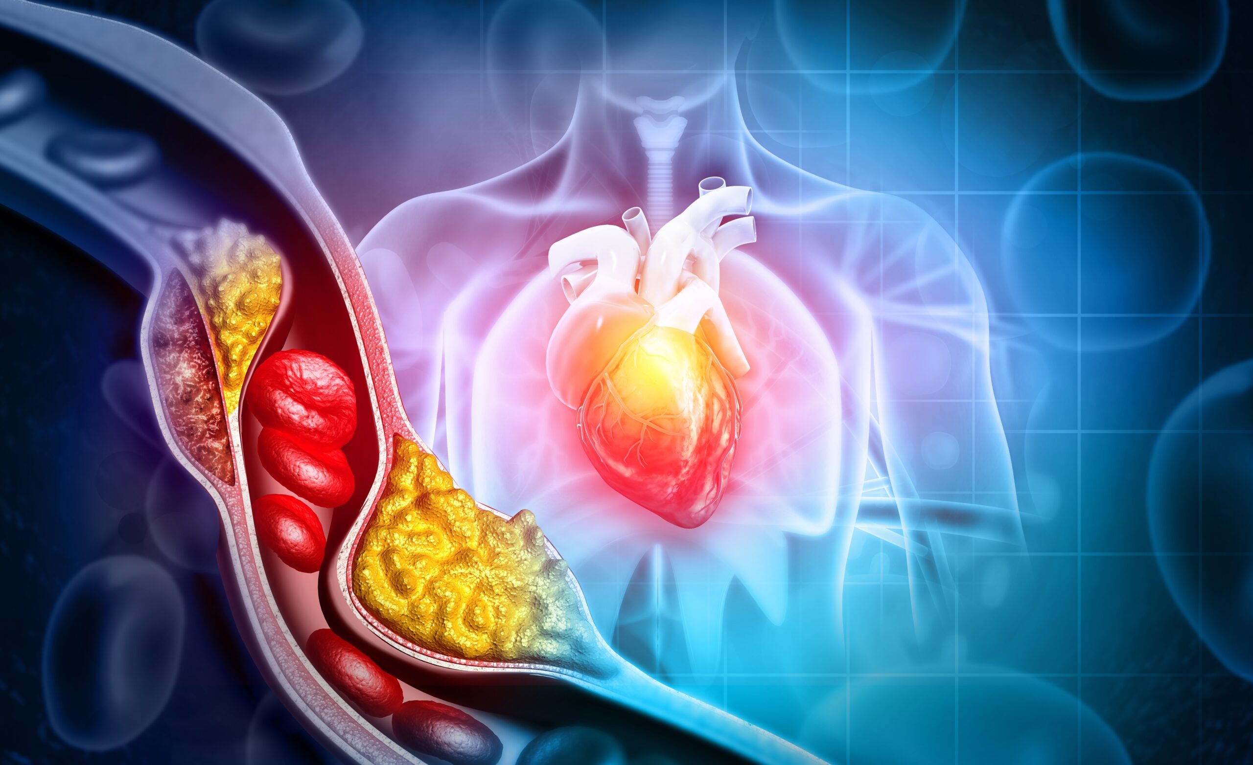
Foods that help lower cholesterol play a crucial role in maintaining heart health and overall well-being. Cholesterol, a waxy substance found in your blood, is essential for building healthy cells. However, high levels of cholesterol can increase the risk of heart disease. This is where diet becomes important, as certain foods can help reduce cholesterol levels.

Oats are a great starting point. A simple change like starting your day with a bowl of oatmeal or oat-based cereal can make a significant difference. Oats contain a type of soluble fiber known as beta-glucan. This fiber helps reduce the absorption of cholesterol into your bloodstream. Including oats in your diet can lower LDL (low-density lipoprotein) or “bad” cholesterol, which is a significant contributor to heart disease.

Another powerful food in the fight against high cholesterol is fish, particularly fatty fish like salmon, mackerel, and sardines. These fish are rich in omega-3 fatty acids, known for their heart-health benefits. Omega-3s help reduce triglycerides in the bloodstream and also slow down the growth rate of atherosclerotic plaque, which can clog arteries. Incorporating fish into your diet a couple of times a week can be beneficial, especially when it replaces sources of saturated fats, like red meat.

Nuts, especially almonds, walnuts, and pistachios, are also beneficial for lowering cholesterol. Nuts contain monounsaturated fats, which are heart-healthy. They also have fiber, which can help lower your LDL cholesterol. Eating a handful of nuts every day can have a positive effect on your heart health. However, nuts are high in calories, so it’s important to eat them in moderation.

Vegetables are an integral part of a cholesterol-lowering diet. They’re high in soluble fiber, which helps lower cholesterol levels. Dark leafy greens, like spinach and kale, contain lutein and other carotenoids, which are associated with a reduced risk of heart disease. Vegetables also have a low calorie-density, meaning you can eat large servings without consuming too many calories, helping in weight management, which is crucial for maintaining healthy cholesterol levels.

Beans are another excellent source of soluble fiber. They also take a while for the body to digest, meaning you feel full for longer after a meal. This is beneficial for weight management. Beans are versatile and can be included in a variety of dishes, making them an easy addition to your diet.

Avocados are a nutrient-dense fruit that can help lower LDL cholesterol. They’re a rich source of monounsaturated fats and fiber, both of which are known for their heart health benefits. Including avocados in your diet can lead to an improvement in your overall cholesterol profile.

Whole grains, like brown rice, barley, and whole wheat, contain more soluble fiber than refined grains. Including more whole grains in your diet can help lower total cholesterol and LDL cholesterol.

Fruits, particularly those high in fiber like apples, grapes, strawberries, and citrus fruits, can also contribute to cholesterol reduction. These fruits are rich in pectin, a type of soluble fiber that lowers LDL cholesterol.

Soy foods, such as tofu and soy milk, can also help lower LDL cholesterol. Studies have shown that consuming soy protein in place of animal protein can lead to a modest reduction in cholesterol levels.

Lastly, dark chocolate and cocoa can also play a role in lowering cholesterol. They contain flavonoids, antioxidants that help lower LDL levels. However, it’s important to choose dark chocolate with a high percentage of cocoa and consume it in moderation due to its high calorie and sugar content.

In conclusion, incorporating a variety of these cholesterol-lowering foods into your diet can significantly impact your overall heart health. It’s also important to remember that lifestyle changes, including regular exercise, not smoking, and maintaining a healthy weight, work in conjunction with a healthy diet to lower cholesterol levels. By making these dietary changes, you can reduce your risk of heart disease and improve your overall health.