Back Pain in Children 

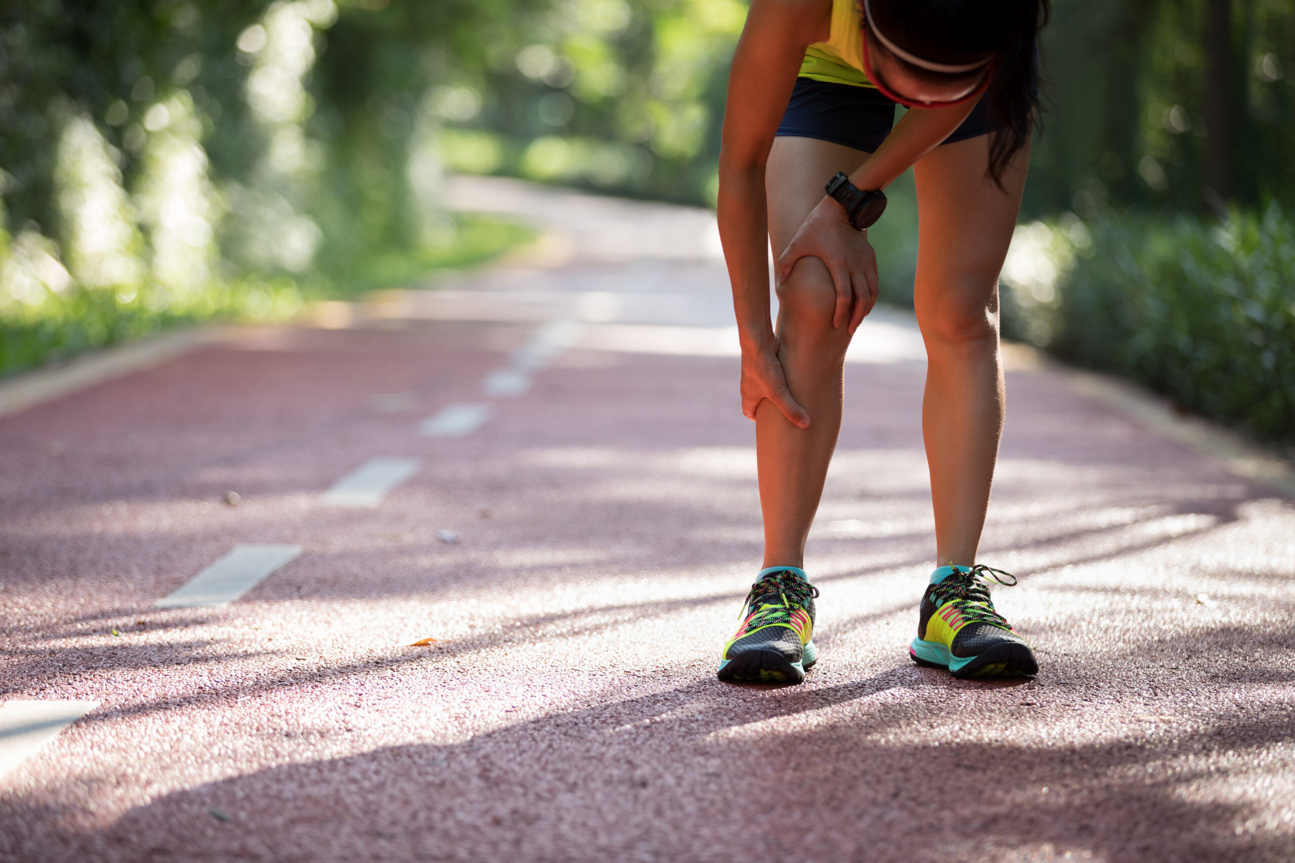
Back pain in children and adolescents occurs more frequently than previously thought (Bernstein, Haus). Incidence depends on age and physical activity, but it may reach up to 24-36% of the pediatric population (Haus). While back pain in adults has been extensively studied, leading to its recognition as a leading cause of disability and a large burden on the healthcare system, back pain in children has received relatively less attention (Bernstein). Current data suggests that it is more likely to be caused by an identifiable, specific underlying cause than for adults; furthermore, some causes of pediatric back pain are serious conditions warranting rapid treatment (AAOS, Bernstein). 

There are several mechanical problems that can arise from physical activity or physical habits, which are often acute and treatable. Haus and Micheli (2012) propose that sports-related injuries contribute to the surprisingly high incidence of pediatric back pain. As is generally the case within the musculoskeletal system, acute injuries or chronic overuse can damage joints and cause pain (AAOS, Haus). The combination of intensive sports training and anatomical changes due to growth can place excessive pressure on the spine and back muscles (Haus). This type of back pain can usually be managed with rest, NSAIDs if needed, and progressive rehabilitation (AAOS, Haus). 

One concern, especially with children, is that heavy backpacks may overload the back. However, there are conflicting points of view – the American Academy of Orthopaedic Surgeons lists carrying a heavy backpack as a cause of muscle strain and imbalance, but a study found no association between heavy backpacks and back pain in school-age children (Haus, AAOS). 

There are also a number of other spinal conditions that can cause back pain in children. While one study found that lumbar strain or spasm was the most common diagnosis given to adolescents with low back pain (at 9%), the next most common were scoliosis (5%), degenerative disc disease (2%), and disc herniation (1%) (Yang). Scoliosis can increase the risk of other back injuries due to imbalanced load during sports. Degenerative disc disease and disc herniation are more common in pediatric athletes than pediatric non-athletes – this population and their parents/guardians should be aware of the risks and consider whether instances of back pain may have underlying pathology instead of being an acute injury (Haus). Other potential causes of pediatric back pain include spondylolysis (stress fracture), spondylolisthesis (slipped disc), and more rarely, infection or tumor (AAOS, Bernstein, Haus, Yang). 

In many cases, pediatric back pain can be managed conservatively with good outcomes. However, there are key factors that should prompt urgent care-seeking: back pain in children aged 4 or younger and back pain with fever, weight loss, nighttime pain, or weakness (AAOS, Berstein, Haus). 

Treatment for children with back pain varies depending on symptoms, diagnosis, and individual-specific factors. Many musculoskeletal injuries and conditions can be managed with a combination of rest, anti-inflammatory medication, and physical therapy. Surgery may be required for some cases of spinal fracture, herniated disc, progressive spondylolisthesis, severe scoliosis, and tumor (Berstein, Haus). Chiropractic management may also be appropriate: a study found that 62% of a group of pediatric patients with low back pain experienced a clinically important decrease in reported pain and 87% perceived a clinically important improvement within a 6-week treatment course. However, patients with chronic low back pain were less likely to respond within the treatment period (Hayden). 

References 

American Academy of Orthopaedic Surgeons. (2019). “Back Pain in Children.” OrthoInfo. https://orthoinfo.aaos.org/en/diseases–conditions/back-pain-in-children/ 

Bernstein, R. M., & Cozen, H. (2007). Evaluation of back pain in children and adolescents. American family physician, 76(11), 1669–1676. 

Haus, B. M., & Micheli, L. J. (2012). Back Pain in the Pediatric and Adolescent Athlete. Clinics in Sports Medicine, 31(3), 423–440. doi:10.1016/j.csm.2012.03.011 

Hayden, J. A., Mior, S. A., & Verhoef, M. J. (2003). Evaluation of chiropractic management of pediatric patients with low back pain: A prospective cohort study. Journal of Manipulative and Physiological Therapeutics, 26(1), 1–8. doi:10.1067/mmt.2003.11 

Yang, S., Werner, B. C., Singla, A., & Abel, M. F. (2017). Low Back Pain in Adolescents. Journal of Pediatric Orthopaedics, 37(5), 344–347. doi:10.1097/bpo.0000000000000653  

Health Insurance Coverage for Chiropractic Care

Chiropractic care is useful as a treatment for many conditions, including low back pain [1]. LBP is one of the leading reasons why Americans visit medical practitioners, as well as a significant contributor to opioid use in the country [1]. Despite chiropractic care’s demonstrated efficacy in helping sufferers of LBP, not all health insurance plans in the United States cover chiropractic care, and those that do tend to do so sparingly [2]. This article will explore differences in chiropractic care coverage across health insurance plans, as well as the adverse effects of limited coverage on patients. 

Most health insurance plans include some coverage for chiropractic care [3]. Heyward et al. conducted a survey of 15 commercial, 15 Medicare Advantage Health, and 15 Medicaid plans [2]. The plans they surveyed represented more than 50% of insured people in the US [2]. On the whole, all three types of plans tend to cover chiropractic care [2]. 

Despite this broad-based coverage, benefits could be subject to certain limits that reduce patient access to chiropractic services [2]. Commercial insurers and Medicaid tend to subject patients to visit limits for chiropractic care [2]. Instead of visit limits, other Medicaid plans may require that beneficiaries receive prior authorization, meet certain conditions, or acquire a referral before visiting a chiropractor [2].  

Meanwhile, Medicare only allows beneficiaries to receive chiropractic coverage if they need to fix a vertebral subluxation [4]. Any other form of chiropractic care, as well as any examinations that the chiropractor orders, will not be covered [4]. This coverage strategy is incoherent, given how chiropractic care for a variety of conditions is associated with reduced opioid-associated disability, lower medical costs, and faster recovery, among many other benefits [5]. 

Costs varied across insurers as well. Medicare Advantage requires beneficiaries to pay a median $20 co-payment for in-network care or a 35% co-insurance fee for out-of-network care [2]. By comparison, commercial insurers place a significantly higher out-of-pocket cost on beneficiaries. Patients pay on average a $60 co-payment per visit if their provider is in-network or a 50% co-insurance fee for out-of-network providers [2]. After meeting their deductible, Medicare beneficiaries pay 20% of the Medicare-Approved Amount for chiropractic services [4]. 

Evidently, there are major deficiencies in chiropractic care coverage across US health insurance plans. Given the many advantages of chiropractic care, these shortcomings are significant. Davis et al. estimated that, for every 1,000 Medicare beneficiaries, reduced access to chiropractic care results in an increased cost of $114,967 [6]. Furthermore, chiropractic care has helped dampen the opioid pandemic by providing patients with nonpharmacological alternatives to back and neck problems [3]. By reducing access to chiropractic care, efforts to combat opioid overuse may be impaired. 

Because of heightened costs, visit limits, and other obstacles, many people who may benefit from chiropractic care cannot access it [1]. To address this issue, researchers recommend pushing for nonpharmacological care, possibly through financial incentives, instructive advocacy, policymaking, or a combination of approaches [1]. By legitimizing nonpharmacological options as frontline treatments for chronic conditions, chiropractic care may become more broadly insured across the US [1]. 

References 

[1] C. M. Goertz and S. Z. George, “Insurer Coverage of Nonpharmacological Treatments for Low Back Pain–Time for a Change,” JAMA Network Open, vol. 1, no. 6, p. 1-3, October 2018. [Online]. Available: DOI: 10.1001/jamanetworkopen.2018.3037

[2] J. Heyward et al., “Coverage of Nonpharmacologic Treatments for Low Back Pain Among US Public and Private Insurers,” JAMA Network Open, vol. 1, no. 6, p. 1-14, October 2018. [Online]. Available: DOI: 10.1001/jamanetworkopen.2018.3044

[3] B. J. Eovaldi and B. McAlpine, “Increased Utilization of Spinal Manipulation by Chiropractors to Tackle the Opioid Epidemic,” Medical Care, vol. 59, no. 12, p. 1039-1041, December 2021. [Online]. Available: DOI: 10.1097/MLR.0000000000001633

[4] “Chiropractic services,” Medicare. [Online]. Available: https://www.medicare.gov/coverage/chiropractic-services

[5] R. A. Leach, “Full-Coverage Chiropractic in Medicare: A Proposal to Eliminate Inequities, Improve Outcomes, and Reduce Health Disparities Without Increasing Overall Program Costs,” Journal of Chiropractic Humanities, vol. 27, no. C, p. 29-36, December 2020. [Online]. Available: DOI: 10.1016/j.echu.2020.10.002

[6] M. Davis et al., “The Effect of Reduced Access to Chiropractic Care on Medical Service Use for Spine Conditions Among Older Adults,” Journal of Manipulative and Physiological Therapeutics, vol. 44, no. 5, p. 353-362, June 2021. [Online]. Available: DOI: 10.1016/j.jmpt.2021.05.002

Low Back Pain: Chiropractic vs. Physical Therapy 

Low back pain (LBP) is a significant burden on health levels and healthcare costs in the United States [1]. Not only can sufferers of LBP experience severe, often disability-inducing pain, but they may also require costly medical care to treat their conditions [1]. Unfortunately, neither the chief cause nor the ideal treatment for LBP is certain [2]. Fortunately, many treatment options for low back pain are available to patients, including chiropractic care and physical therapy (PT) [2]. When deciding whether to opt for one over another, there are a few key characteristics and discoveries that patients and their medical teams should keep in mind. 

Chiropractic care consists of a wide array of treatments, including electrical stimulation, heat or ice application, and, most commonly, joint manipulations for the correction of subluxations [3]. In the context of LBP treatment, there has been disagreement among experts about the appropriate role of chiropractic care [2]. 

Some of this debate may have been quelled by a 2018 study [2]. The study sought to analyze the efficacy of chiropractic in treating LBP [2]. The researchers compared how active-duty military personnel complaining of LBP fared with usual care alone (ranging from pharmaceutics to PT) versus usual care combined with up to twelve chiropractic treatments [2]. At the end of the six-week treatment period, the usual care plus chiropractic group reported less severe pain, reduced disability, diminished pain medication use, improved function, and higher satisfaction [2]. According to these results, chiropractic appears to be an effective treatment for LBP.  

Similarly, physical therapy has reportedly contributed to improved outcomes when compared to no treatment or medical intervention alone [1]. The central objective of PT is to prevent further disability and improve functional capability [4]. For combating low back pain, the most effective forms of physical therapy appear to be active strategies, such as yoga, tai chi, and other forms of exercise [4]. Because no single form of PT has emerged as superior to all others, researchers suggest that patients employ a diversity of techniques, chosen according to personal preference [4]. 

Multiple studies have compared chiropractic care and PT’s respective effects on LBP. For instance, a 2006 UCLA study focused on the differences in disability, remission, and pain intensity among LBP patients sorted into four groups: chiropractic with and without physical modalities, and medical care with and without PT [5]. After 18 months of analyzing their subjects, Hurwitz et al. found that the groups were relatively equivalent across the primary outcomes [5]. However, the chiropractic group had a “greater likelihood of perceived improvement,” suggesting that this technique may be associated with a boost in confidence [5]. 

Further evidence suggests that each method may have unique benefits. Gudavalli and colleagues compared flexion-distraction (FD), a chiropractic technique, with active trunk exercise protocol (ATEP), a form of PT [6]. The FD patients experienced greater pain relief, leading the researchers to conclude that FD may be more appropriate for patients with chronic LBP or those with radiculopathy [6]. Meanwhile, patients with recurrent pain improved most under ATEP [6].   

Besides the severity of the patient’s condition, cost may also influence which treatment they choose. A 2020 study found that the total average cost for LBP patients who opted for chiropractic treatment was $48.56 lower than those who chose physical therapy [1]. 

Nevertheless, there is no ideal treatment for low back pain. However, by considering their degree of pain, their desire for extra motivation, and budget concerns, patients can make a more informed choice between physical therapy and chiropractic care. 

References 

[1] N. Khodakarami, “Treatment of Patients with Low Back Pain: A Comparison of Physical Therapy and Chiropractic Manipulation,” Healthcare, vol. 8, no. 44, p. 1-8, February 2020. [Online]. Available: https://doi.org/10.3390/healthcare8010044

[2] R. H. Shmerling, “Should you see a chiropractor for low back pain?,” Harvard Health Publishing, Updated August 16, 2019. [Online]. Available: https://www.health.harvard.edu/blog/should-you-see-a-chiropractor-for-low-back-pain-2019073017412

[3] B. Sears, “Here’s How Chiropractors and Physical Therapists Are Different,” VeryWell Health, Updated September 3, 2021. [Online]. Available: https://www.verywellhealth.com/chiropractor-vs-physical-therapy-5194093

[4] E. A. Shipton, “Physical Therapy Approaches in the Treatment of Low Back Pain,” Pain and Therapy, vol. 7, no. 2, p. 127-137, September 2018. [Online]. Available: https://doi.org/10.1007/s40122-018-0105-x

[5] E. L. Hurwitz et al., “A Randomized Trial of Chiropractic and Medical Care for Patients With Low Back Pain: Eighteen-Month Follow-up Outcomes From the UCLA Low Back Pain Study,” Spine, vol. 31, no. 6, p. 611-621, March 2006. [Online]. Available: https://doi.org/10.1097/01.brs.0000202559.41193.b2

[6] M. R. Gudavalli et al., “A randomized clinical trial and subgroup analysis to compare flexion–distraction with active exercise for chronic low back pain,” European Spine Journal, vol. 15, no. 7, p. 1070-1082, December 2005. [Online]. Available: https://doi.org/10.1007/s00586-005-0021-8.

Health Risks of Trans Fats 

Trans fats have broadly been recognized as an unhealthy form of fat that is present in the diets of many people, especially in the U.S. Though it is true that almost any nutrient in excess can be harmful, when it comes to fat, trans fats are associated with the worst health risks. Dietary fat can be grouped into four categories based on characteristics of their molecular structure that influence how they affect the body, in order of most to least healthy: polyunsaturated, monounsaturated, saturated, and trans (1). Extensive research has established clear recommendations to avoid the latter two types and/or replace with the first two types when possible (1-3). However, it is important to note that fats are a necessary nutrient for normal cellular functioning (1). 

The advantages of trans fats are that they are inexpensive to produce and store well, making them attractive for food preparation at both the commercial and individual level (2). Trans fats (a.k.a. partially hydrogenated oils) can be produced artificially from liquid oil, which typically contains mostly unsaturated fats, by changing its molecular structure and turning it into a solid (2-4).  

Specifically, unsaturated fats are hydrogenated – the fatty acid chains of unsaturated fats contain one or more cis double bonds, causing “kinks” that make it more difficult for the substance to pack in an orderly manner into a solid. Hydrogenation causes some of those double bonds to become single bonds and some to become trans double bonds, both of which straighten out the molecule’s structure and increase the substance’s melting point. 

Research in the past few decades led to the discovery of the many health risks of trans fats. In particular, these fats increase LDL cholesterol and decrease HDL cholesterol, which are respectively considered the “bad” and “good” kind (1-3). They are known to increase the risk of heart disease and stroke, and are also associated with a higher risk of type 2 diabetes (2,3). Consuming 5 grams of trans fats each day increases your risk of ischemic heart disease by 25% (5). Monkeys that were fed a diet with trans fats over six years gained weight, had increased fat within the abdomen, and showed signs of poorer glucose regulation compared to the control group who received the equivalent diet with unsaturated fats (4). 

Fortunately, restrictions on this type of fat are tightening worldwide. Denmark, for example, has strongly restricted the use of trans fats in all food products, and a comparison across twenty countries found that products from the same fast-food chains had much less trans fat in Denmark (1,5). Several jurisdictions in the U.S. have also implemented restrictions, while the FDA requires trans fats to be identified in ingredient and nutritional food labels nationwide.  

There are also easy steps that U.S. residents can take to monitor their fat intake, such as looking for the amount of trans fat in a nutritional label or for “partially hydrogenated oils” in the ingredients label (2-4). Furthermore, most fried foods and many baked treats contain high levels of trans fat (2,3). Solid vegetable shortening and margarine should be avoided as much as possible (2-4); note that butter, while solid, is made from milk fat in a different process and contains high levels of saturated fat but relatively small amounts of trans fat. Unsaturated fats are relatively healthier, though they should still be consumed in moderation, and include canola, safflower, and olive oil (non-tropical plant oils) (1-3). 

References 

  1. AHA. “Dietary Fats.” American Heart Association. Reviewed November 1, 2021. https://www.heart.org/en/healthy-living/healthy-eating/eat-smart/fats/dietary-fats 
  1. AHA. “Trans Fats.” American Heart Association. Reviewed March 23, 2017. https://www.heart.org/en/healthy-living/healthy-eating/eat-smart/fats/trans-fat 
  1. Bridges M. “Facts about trans fats.” MedlinePlus. Updated May 26, 2020. https://medlineplus.gov/ency/patientinstructions/000786.htm 
  1. Stender S, Dyerberg J, Astrup A. High Levels of Industrially Produced Trans Fat in Popular Fast Foods. NEJM. 2006;354(15):1650-1652. doi: 10.1056/nejmc052959 
  1. Kavanagh K, Jones KL, Sawyer J, Kelley K, Carr JJ, Wagner JD, Rudel LL. Trans Fat Diet Induces Abdominal Obesity and Changes in Insulin Sensitivity in Monkeys. 2012;15(7):1675-1684. doi: 10.1038/oby.2007.200

Natural Detoxification Processes 

Toxins are substances produced by living organisms which are harmful to humans. For example, certain natural toxins are often part of a plant’s defense mechanisms and can thus be found in foods. Food may also contain toxins as a result of containing toxin-producing mold or microorganisms [1]. Other toxins may derive from bacteria; cholera’s clinical manifestations, for example, result from a toxin produced by cholera bacteria [2]. In fact, many people consume alcohol, a toxin, for effects that are related to its toxicity. Fortunately, the human body has a number of natural detoxification processes that help protect health.  

The terms “toxins” and “toxicants”, which are the synthetic equivalents of toxins, are sometimes used interchangeably. Toxicants may include environmental chemicals or medicinal products that are poisonous in large amounts. The latter includes but is not limited to lead, pesticides, flame retardants, and polychlorinated biphenyls. 

Detoxification corresponds to the physiological removal of such toxins and toxicants from a living organism, such as the human body. The liver and kidney are naturally capable of detoxification, as are intracellular proteins such as cytochrome (CYP) enzymes present in mitochondria or the endoplasmic reticulum. 

The liver plays a critical role in protecting our bodies from potentially toxic chemicals by converting lipophilic elements into more water-soluble metabolites which can be eliminated through urine. The substrate specificity, abundance of isoenzymes, and inducibility of these enzyme systems present in the liver make them well-suited to metabolize the many different types of substances that we are exposed to [3].  

The kidneys, meanwhile, are responsible for filtering blood and removing waste, acid, and extra fluid from the body. They are key to maintaining a healthy equilibrium of water, salts, and minerals. However, they require healthy habits to operate at maximum capacity: Drinking plentiful water and eating kidney-friendly foods (vegetables, whole grains, fish, and fruits) help support kidney detoxification [4]. 

Mammalian CYP enzymes can oxidize both xenobiotics and endogenous compounds, and are key to detoxifying foreign substances, as well as controlling hormone balance, vitamin D metabolism, and cholesterol synthesis. These enzymes are responsible for metabolizing a wide variety of clinically, physiologically, and toxicologically important substances [5].  

Sweating, however, has not been shown conclusively to have a clear detoxifying effect on the body. Humans primarily sweat to cool down rather than to excrete toxic substances. Since many persistent organic pollutants are lipophilic, they do not dissolve in sweat, which consists primarily of water. Even when one does sweat out a certain amount of environmental pollutants, the volumes are minuscule [6]. However, given their greater hydrophilicity, certain amounts of heavy metals and plastics (such as bisphenol A) do dissolve into and are excreted along with sweat to a certain degree.  

A few key habits are critical to maximizing the body’s natural detoxification processes [7]. Individuals should first cease smoking and minimize their consumption of alcohol, both of which add toxins into the body. In addition, individuals need to stay hydrated, eat a nutrient-dense diet [8], and obtain regular and sufficient amounts of sleep so that detoxification processes can function at an optimal level. Finally, maintaining excellent indoor air quality is critical to minimizing one’s exposure to any environmental toxins in the first place. Boosting the body’s own natural detoxification processes is critical to ensuring maximal health and well-being, and can be done with small changes in behavior. 

References 

1. Natural toxins in food. Available at: https://www.who.int/news-room/fact-sheets/detail/natural-toxins-in-food

2. Toxins: MedlinePlus Medical Encyclopedia. Available at: https://medlineplus.gov/ency/article/002331.htm

3. Grant, D. M. Detoxification pathways in the liver. J. Inherit. Metab. Dis. (1991). doi:10.1007/BF01797915 

4. Kidney Detox: Is It Necessary & How To Do It Safely | mindbodygreen. Available at: https://www.mindbodygreen.com/articles/how-to-do-a-kidney-detox.  

5. Esteves, F., Rueff, J. & Kranendonk, M. The Central Role of Cytochrome P450 in Xenobiotic Metabolism—A Brief Review on a Fascinating Enzyme Family. J. Xenobiotics (2021). doi:10.3390/jox11030007 

6. Imbeault, P., Ravanelli, N. & Chevrier, J. Can POPs be substantially popped out through sweat? Environment International (2018). doi:10.1016/j.envint.2017.11.023 

7. Full body detox: How to help the body detox at home. Available at: https://www.medicalnewstoday.com/articles/full-body-detox#contacting-a-doctor. 

8. Klein, A. V. & Kiat, H. Detox diets for toxin elimination and weight management: A critical review of the evidence. J. Hum. Nutr. Diet. 28, 675–686 (2015). doi: 10.1111/jhn.12286 

Prediabetes: Diagnosis, Diet, and Exercise 

Prediabetes is defined as a condition high risk for developing diabetes. Patient blood sugar levels are higher than normal but not high enough to classify a patient as diabetic. The prevalence of prediabetes is increasing and poses a serious public health problem. However, prediabetes is reversible, and proper diagnosis and management can delay or prevent the onset of diabetes (Mainous et al., 2016). 11.3% of the adult US population has diabetes, and 38% of the adult US population has prediabetes (“National Diabetes Statistics Report”, 2022). While medications also play a role, first line treatment of prediabetes primarily includes lifestyle modifications such as diet and exercise.  

Diagnosis of prediabetes often occurs during testing for diabetes. Some patients with prediabetes may experience symptoms, but not all will. Patients with prediabetes should be tested for type 2 diabetes mellitus every one or two years (“Myths about Diabetes”, 2022). Testing involves a 2-hour oral glucose tolerance test which demonstrates impaired fasting glucose and/or an impaired glucose tolerance. Impaired fasting glucose is generally defined as a fasting plasma glucose between 100.8-124.2 mg/dL, and impaired glucose tolerance is defined as plasma glucose values ranging from 140.4-198.0 mg/dL. HbA1c, known as glycosylated hemoglobin, within the rage of 5.7% to 6.4% can also be used to identify individuals with prediabetes (Mainous et al., 2016).  

Current guidelines from the American Diabetes Association state that a healthy meal plan for someone with diabetes is the same healthy eating plan for anyone. This means including nutritious foods such as non-starchy vegetables, limiting added sugars, and prioritizing whole foods when possible (“Myths about Diabetes”, 2022). Reducing caloric intake can lead to an improvement in glycemic control though it is not necessarily associated with weight loss (Sénéchal et al., 2014). During a randomized controlled trial with 34 diabetes/pre-diabetes participants, researchers found that a low carbohydrate diet was more effective at reducing HbA1c values at three months compared to a moderate carbohydrate diet (Saslow et al., 2014). Patients with prediabetes/diabetes do not need to completely avoid carbs, but starchy and high-carb foods will raise their blood sugar. One concern is that a low carbohydrate diet results in a higher proportion of calories from fat, which may also be a cause of concern, but researchers did not find significant elevations in LDL with this diet (Saslow et al., 2014). Continuing research should aim to understand how modifying other macronutrients and micronutrients affects blood sugar levels (Sénéchal et al., 2014).  

Exercise interventions are also essential for controlling prediabetes. Aerobic exercise is known to have transient effects on postprandial glucose metabolism and skeletal muscle insulin sensitivity (Rynders et al., 2014). Generally, a combination of aerobic and resistance training is recommended for prediabetic control (Sénéchal et al., 2014). One study found that although moderate intensity exercise and high intensity exercise did not have significant differences in improving postprandial insulin sensitivity, high intensity interval exercise may have greater postprandial effects compared to moderate exercise (Rynders et al., 2014). Further research is required to investigate the independent effect of exercise on glycemic control in individuals with prediabetes and understand how to optimize the effects of exercise (Sénéchal et al., 2014). 

Ultimately, while it is generally known that dietary and exercise interventions have a significant effect on prediabetes, further research should focus on understanding the independent effects of diet and exercise on individuals with prediabetes. Currently, most patients with prediabetes do not receive adequate care in managing their condition (Mainous et al., 2016). Primary care providers have a large role to play in the prevention of prediabetes/diabetes, and they must be equipped with the proper knowledge and skills to counsel patients.  

References 

Mainous AG 3rd, Tanner RJ, Baker R. Prediabetes Diagnosis and Treatment in Primary Care. J Am Board Fam Med. 2016;29(2):283-285. doi:10.3122/jabfm.2016.02.150252 

Myths about Diabetes. American Diabetes Association. https://www.diabetes.org/tools-support/diabetes-prevention. 

National Diabetes Statistics Report. Centers for Disease Control and Prevention. https://www.cdc.gov/diabetes/data/statistics-report/index.html.

Rynders CA, Weltman JY, Jiang B, et al. Effects of exercise intensity on postprandial improvement in glucose disposal and insulin sensitivity in prediabetic adults. J Clin Endocrinol Metab. 2014;99(1):220-228. doi:10.1210/jc.2013-2687 

Saslow LR, Kim S, Daubenmier JJ, et al. A randomized pilot trial of a moderate carbohydrate diet compared to a very low carbohydrate diet in overweight or obese individuals with type 2 diabetes mellitus or prediabetes. PLoS One. 2014;9(4):e91027. Published 2014 Apr 9. doi:10.1371/journal.pone.0091027 

Sénéchal M, Slaght J, Bharti N, Bouchard DR. Independent and combined effect of diet and exercise in adults with prediabetes. Diabetes Metab Syndr Obes. 2014;7:521-529. Published 2014 Oct 31. doi:10.2147/DMSO.S62367

Diet and Mood Disorders 

The connection between nutrition and mental illnesses of mood is less acknowledged than the negative effect of nutritional deficiencies on physical health. In recent years, nutritional neuropsychology has indicated nutrition is inevitably intertwined with human emotion, behavior, and mood. The newly established International Society for Nutrition Psychiatry Research (ISNPR) aims to understand the relationship between nutrition, metabolism and neuropsychiatric disorders [1]. Epidemiological studies have indicated an unhealthy diet (for example, a meal regimen revolving around fast-food or refined sugar) can increase a person’s risk of depression, while a balanced, healthy diet can protect against such neuropsychiatric disorders [2,3]. 

Macromolecules such as carbohydrates are known to release insulin, which increases the uptake of most amino acids into peripheral tissues, such as muscles. Tryptophan, an amino acid usually present at low levels in the bloodstream regardless of diet, is unaffected by insulin; thus with the secretion of insulin, the ratio of tryptophan to other amino acids drastically increases [4]. Without the competition of other amino acids, tryptophan is rapidly transported across the blood-brain barrier, where it enters the central nervous system and is converted into serotonin [4]. Increases in this “feel-good hormone” is the reason why diets low in carbohydrates often precipitate anhedonic conditions such as seasonal affective disorder (SAD), premenstrual syndrome (PMS), or major depressive disorder (MDD) [4,5]. The glycemic index (GI) is a caveat of this linear association which reveals fruits and vegetables (which have a low GI) tend to provide more long-lasting positive effects on brain chemistry and mood, while the reverse is true for high GI foods, for example processed foods or those high in artificial sugar [5]. 

Many neurotransmitters, which are signal molecules that allow for “communication” between neurons, are derivatives of amino acids. Among amino acids, 12 are made in the body and 8 are acquired from dietary intake. For example, the neurotransmitters dopamine and serotonin, both of which are related to emotional well-being, are synthesized from the amino acids tyrosine and tryptophan, respectively [5]. The extracellular-regulated protein kinase (ERK) pathway has been associated with protective processes such as plasticity and resilience in neuropsychiatric disorders including bipolar disorder and MDD [6]. Using Western blot analyses, researchers found lower levels of proteins within the ERK cascade in the frontal cortex of individuals with mood disorders compared to healthy controls [6]. In a murine model, stimulation of the ERK pathway was induced through administration of lithium or valproate, two commonly used mood stabilizers in the treatment of mood disorders [6]. 

Half of neural gray matter is composed of fatty acids that must be supplied through diet. Omega-6 and omega-3 polyunsaturated fatty acids (PUFAs) are critical in regulating the biochemical aspects of cellular membranes [5]. In a 4-month, double-blind, placebo-controlled study of 30 individuals with bipolar disorder, omega-3 fatty acids improved the course of illness indicated by a significantly longer period of remission for individuals receiving omega-3 treatment compared to placebo [7]. The authors of this study speculated that since omega-3 fatty acids were known to become highly incorporated in neuronal membranes, they could inhibit signal transduction mechanisms in the human central nervous system, much like many pharmacological mood stabilizers [7]. 

In addition, active areas of research include the effect of various vitamins and minerals on mental health and mood disorders. Although certainly many caveats between diet and mood disorders remain, the foundations of nutritional neuroscience provide a preliminary basis for future, more nuanced research, and current information demonstrates the importance of a balanced diet for all aspects of health.

References  

1. ISNPR 2019. (n.d.). International Society for Nutritional Psychiatry Research (ISNPR). Retrieved from http://www.isnpr.org/isnpr2019/  

2. Sánchez-Villegas, A., Toledo, E., Irala, J. de, Ruiz-Canela, M., Pla-Vidal, J., & Martínez-González, M. A. (2012). Fast-food and commercial baked goods consumption and the risk of depression. Public Health Nutrition, 15(3), 424–432. https://doi.org/10.1017/S1368980011001856  

3. Jacka, F. N., O’Neil, A., Opie, R., Itsiopoulos, C., Cotton, S., Mohebbi, M., Castle, D., Dash, S., Mihalopoulos, C., Chatterton, M. L., Brazionis, L., Dean, O. M., Hodge, A. M., & Berk, M. (2017). A randomised controlled trial of dietary improvement for adults with major depression (The ‘smiles’ trial). BMC Medicine, 15(1), 23. https://doi.org/10.1186/s12916-017-0791-y  

4. Wurtman, R. J., & Wurtman, J. J. (1989). Carbohydrates and depression. Scientific American, 260(1), 68–75. https://www.jstor.org/stable/24987109 

5. Rao, T. S. S., Asha, M. R., Ramesh, B. N., & Rao, K. S. J. (2008). Understanding nutrition, depression and mental illnesses. Indian Journal of Psychiatry, 50(2), 77–82. https://doi.org/10.4103/0019-5545.42391 

6. Yuan, P., Zhou, R., Wang, Y., Li, X., Li, J., Chen, G., Guitart, X., & Manji, H. K. (2010). Altered levels of extracellular signal-regulated kinase signaling proteins in postmortem frontal cortex of individuals with mood disorders and schizophrenia. Journal of Affective Disorders, 124(1), 164–169. https://doi.org/10.1016/j.jad.2009.10.017 

7. Stoll, A. L., Severus, W. E., Freeman, M. P., Rueter, S., Zboyan, H. A., Diamond, E., Cress, K. K., & Marangell, L. B. (1999). Omega 3 fatty acids in bipolar disorder: A preliminary double-blind, placebo-controlled trial. Archives of General Psychiatry, 56(5), 407–412. https://doi.org/10.1001/archpsyc.56.5.407 

Supplements for Joint Cartilage Health

            Cartilage is the fibrous and elastic connective tissue that exists throughout the body and serves various purposes.1 Joint cartilage, known as articular cartilage, covers bones in locations such as the knee, hip, and wrist, allowing the bones in a joint to smoothly glide over one another and absorb shock.1 Cartilage damage is a common injury that may occur from a sudden injury or from gradual wear and tear,2 the latter of which can lead to osteoarthritis, a common form of arthritis characterized by pain, stiffness, and swelling in the joints resulting from the partial or complete breakdown of joint cartilage.3  Cartilage health is a significant public health issue: over 90 million Americans suffer from osteoarthritis alone.4 Many different approaches to support cartilage health exist, ranging from physical activity to dietary supplements.

            Cartilage, which lacks blood vessels, does not heal well on its own. While more severe cases of cartilage damage may require replacement of the entire joint or other surgical intervention, self-care measures exist for moderate cases. Supplements are a popular form of such treatment that have been found to alleviate joint pain and reduce joint inflammation. Specifically, the supplements glucosamine (GlcN) and chondroitin sulfate (CS) have been the subject of many studies, most of which corroborate their efficacy in improving cartilage health but some of which were inconclusive.

            GlcN is a type of glycosaminoglycan (a polysaccharide containing amino groups) that exists naturally in cartilage and that can delay degeneration and reduce osteoarthritic pain when administered orally.5 GlcN has multiple properties that enable it to achieve these physiological effects. Its primary mechanism of action is to reduce the inflammation associated with OA; it lowers the expression of p38 mitogen-activated protein kinase (MAPK) and c-Jun N-terminal kinase (JNK), factors associated with OA-related inflammation.6 GlcN also has antioxidant properties. Like the antioxidants curcumin and vitamin E, GlcN can alleviate pain and restore joint function in OA patients.7 There are several possible mechanisms for this effect, including the ability of GlcN to upregulate antioxidant enzyme levels and to suppress levels of damaging oxygen radicals.8 However, some studies have shown that glucosamine did not improve OA: Kwoh et al. studied the knee joint structure of those with chronic knee pain using magnetic resonance imaging and found no change after a 24-week GlcN treatment.9 Results such as these demonstrate the need for further studies on the efficacy of GlcN.

            Like GlcN, CS is also a natural component of cartilage and is in part responsible for its resistance and elasticity.10 In addition to its anti-inflammatory effect, CS also resolves the symptoms of OA by increasing the synthesis of type II collagen and proteoglycan, components of cartilage, in chondrocytes.11 CS has anti-apoptic effects; it has been found to reduce the number of apoptosis events in vitro and in vivo.12

            S-adenosylmethionine (SAMe) is another compound among supplements for OA that, like the first two, has largely been found to be effective in improving cartilage health. SAMe is synthesized from methionine and ATP and is involved in several biochemical pathways.13 A possible mechanism of action unique to SAMe relates to its observed effect on the liver. Alcoholic liver disease can arise from decreased SAMe levels because SAMe functions to reduce oxidative stress.13 This same phenomenon may occur in the joints, which could explain why taking SAMe as a supplement helps reduce joint pain.

References

1. MD, E. K. What Is Cartilage? Arthritis Health https://www.arthritis-health.com/types/joint-anatomy/what-cartilage.

2. Cartilage damage. National Health Service https://www.nhs.uk/conditions/cartilage-damage/ (2017).

3. Osteoarthritis (OA) | Arthritis. CDC. https://www.cdc.gov/arthritis/basics/osteoarthritis.htm (2020).

4. Krishnan, Y. & Grodzinsky, A. J. Cartilage Diseases. Matrix Biol. J. Int. Soc. Matrix Biol. 71–72, 51–69 (2018). doi:10.1016/j.matbio.2018.05.005.

5. Al-Saadi, H. M., Pang, K.-L., Ima-Nirwana, S. & Chin, K.-Y. Multifaceted Protective Role of Glucosamine against Osteoarthritis: Review of Its Molecular Mechanisms. Sci. Pharm. 87, 34 (2019).

6. Wen, Z.-H. et al. Glucosamine sulfate reduces experimental osteoarthritis and nociception in rats: association with changes of mitogen-activated protein kinase in chondrocytes. Osteoarthritis Cartilage 18, 1192–1202 (2010).

7. Cheng, D. W. et al. An analysis of high glucose and glucosamine-induced gene expression and oxidative stress in renal mesangial cells. Arch. Physiol. Biochem. 112, 189–218 (2006).

8. Mendis, E., Kim, M.-M., Rajapakse, N. & Kim, S.-K. Sulfated glucosamine inhibits oxidation of biomolecules in cells via a mechanism involving intracellular free radical scavenging. Eur. J. Pharmacol. 579, 74–85 (2008).

9. Kwoh, C. K. et al. Effect of Oral Glucosamine on Joint Structure in Individuals With Chronic Knee Pain: A Randomized, Placebo-Controlled Clinical Trial. Arthritis Rheumatol. 66, 930–939 (2014).

10. Henrotin, Y., Mathy, M., Sanchez, C. & Lambert, C. Chondroitin Sulfate in the Treatment of Osteoarthritis: From in Vitro Studies to Clinical Recommendations. Ther. Adv. Musculoskelet. Dis. 2, 335–348 (2010).

11. Wang, L., Wang, J., Almqvist, K. F., Veys, E. M. & Verbruggen, G. Influence of polysulphated polysaccharides and hydrocortisone on the extracellular matrix metabolism of human articular chondrocytes in vitro. Clin. Exp. Rheumatol. 20, 669–676 (2002).

12. Jomphe, C. et al. Chondroitin sulfate inhibits the nuclear translocation of nuclear factor-kappaB in interleukin-1beta-stimulated chondrocytes. Basic Clin. Pharmacol. Toxicol. 102, 59–65 (2008).

13. Hosea Blewett, H. J. Exploring the mechanisms behind S-adenosylmethionine (SAMe) in the treatment of osteoarthritis. Crit. Rev. Food Sci. Nutr. 48, 458–463 (2008).

Health Effects of Blue Light

As work, school, and much of daily life become increasingly dependent on computers, concerns about the effect of screen use on sleep and brain functioning grow in the scientific community. Some studies indicate that exposure to light at night, especially blue light, may be linked to chronic health conditions such as diabetes, heart disease, and some cancers.1–3 In a Harvard study, researchers shifted the sleep schedules of participants to alter their circadian rhythms and noticed that their blood sugar levels increased while their levels of leptin, a hormone that regulates the feeling of fullness, decreased.4 Research on the precise health effects of blue light at night is inconclusive as high screen use is often correlated with sedentary behavior and poor sleep hygiene, both of which are proven to be detrimental to human health.  

Light plays an integral role in human circadian responses, regulating sleep and alertness by suppressing release of the “sleep hormone,” melatonin. About 20 years ago, scientists discovered that the human eye contains not only visual photoreceptors for detecting changes in color and light, but also photosensitive retinal ganglion cells that trigger circadian responses.3 These cells are particularly sensitive to blue light, likely because these wavelengths of light (415-455 nm) are also found in the sky. During the day, exposure to blue light is associated with increased attention, mood, and reaction times, but at night, it suppresses melatonin and shifts circadian rhythms more than any other color of light.4  

Concentrated blue light may also have a direct effect on the retina of the eye, causing photochemical injury. Blue light contains higher energy than most other colors, and long term exposure can cause visual fatigue and nearsightedness.5 Increased exposure to sunlight, which is primarily composed of blue light, is a risk factor for cataract formation.  

A study published in 2020 found that a third of study participants used a blue-light emitting device 9 to 11 hours a day and another 16% used the devices 12 to 14 hours per day.6 Despite inconclusive research on the links between blue light and physical conditions, higher screen time is associated with moderate to severe depression.7 The implications of this research are particularly concerning for children and adolescents, who are more susceptible to fluctuations in circadian rhythms and negative effects of light on brain functioning and academic performance.  

There are several methods to ameliorate or diminish the effects of blue light, but the best solution is to reduce screen time at night. Scientists suggest using dim red lights for night lights, as red wavelengths are less likely to alter circadian rhythms and suppress melatonin production. For computers and phones, there are free apps that filter blue and green lights at night, including f.lux and Twilight. Blue light glasses may also be useful–a University of Toronto study found that participants wearing blue light glasses when exposed to bright light had similar melatonin levels as participants in dark environments at night.3 Many LED lights, while more environmentally friendly than their incandescent counterparts, emit more blue light than traditional bulbs that give off significantly more heat than light. As LED lights are relatively new, there are no long-term studies about the effects of LEDs over the course of the human lifespan. To reduce the emission of blue light, there are films and covers for LED bulbs that can make them warmer. Finally, spending time outside during the day can restore natural circadian rhythms and reduce unusual sleep patterns. Blue light may be necessary for much of daily life, but it is possible to reduce potential side effects of blue light, and it may be helpful to limit screen use outside of work and school.  

References 

1. Cho Y, Ryu SH, Lee BR, Kim KH, Lee E, Choi J. Effects of artificial light at night on human health: A literature review of observational and experimental studies applied to exposure assessment. Chronobiology International. 2015;32(9):1294-1310. doi:10.3109/07420528.2015.1073158 

2. Opperhuizen AL, Stenvers DJ, Jansen RD, Foppen E, Fliers E, Kalsbeek A. Light at night acutely impairs glucose tolerance in a time-, intensity- and wavelength-dependent manner in rats. Diabetologia. 2017;60(7):1333-1343. doi:10.1007/s00125-017-4262-y 

3. Fleming A. The truth about blue light: does it really cause insomnia and increased risk of cancer?https://www.theguardian.com/lifeandstyle/2018/may/28/blue-light-led-screens-cancer-insomnia-health-issues. Published May 28, 2018.  

4. Harvard Health Publishing. Blue light has a dark side: What is blue light? The effect blue light has on your sleep and more. Harvard Medical School. https://www.health.harvard.edu/staying-healthy/blue-light-has-a-dark-side. Published July 7, 2020. 

5. Li H, Zhang M, Wang D, et al. Blue Light from Cell Phones Can Cause Chronic Retinal Light Injury: The Evidence from a Clinical Observational Study and a SD Rat Model. Koike C, ed. BioMed Research International. 2021;2021:1-13. doi:10.1155/2021/3236892 

6. Bahkir F, Grandee S. Impact of the COVID-19 lockdown on digital device-related ocular health. Indian J Ophthalmol. 2020;68(11):2378. doi:10.4103/ijo.IJO_2306_20 

7. Madhav KC, Sherchand SP, Sherchan S. Association between screen time and depression among US adults. Preventive Medicine Reports. 2017;8:67-71. doi:10.1016/j.pmedr.2017.08.005 

How Orthotics Correct Posture

Orthotics are assistive technologies that can address many musculoskeletal concerns by correcting position and/or supporting proper function [1]. The most common type is the shoe insert. Since the rise of rapid prototyping techniques, orthotics have become more personalized to individual patients and thus more effective than ever [1]. A central function of orthotics is to correct posture [1].

In a case study involving a 59-year-old male patient with ankylosing spondylitis (AS), an orthotic device was able to alleviate some of the patient’s postural problems and, thereby, postpone his later surgical treatment [2]. The patient received orthotic devices to treat his postural imbalance [2]. Within two weeks of wearing a 9-millimeter gel heel lift on one side, the patient’s posture had markedly improved [2]. The researchers believed that this successful correction could be explained by the heel lift leveling the patient’s hip posture so that he no longer had to walk on his tiptoes [2]. Although this study is but a single investigation into heel lift orthotics, it suggests that orthotics can promote better posture by leveling out patients’ gaits to correct any disruptive asymmetries.

A different study focused on the efficacy of plantar orthotics in promoting improved posture [3]. Foot health is crucial for correct posture because of the foot’s key role for support, weight transfer, and propulsion [3]. In this study, the device in question was an insole technology known as Regular Gait that supports the plantar arch [3]. The experimenters monitored 30 subjects as they wore the RG insoles [3]. None of the subjects suffered from any foot diseases or damage [3]. The orthotics stimulated plantar mechanoreceptors, which promoted a reorganization of users’ plantar stance up to the higher nervous system [3]. By influencing the kinetic biofeedback chains that originate at the level of the foot and travel up to the brain, plantar insole orthotics can significantly improve their users’ upright posture [3].

Another means by which orthotics can correct posture is by beneficially arranging limbs during sleep [4]. This was the case in a study following a patient who suffered from spastic diplegic cerebral palsy [4]. A defining characteristic of the condition is increased muscle tone, which can lead patients to adopt incorrect postures [4]. By arranging the patient’s knee in a way that would improve his posture while he was asleep, the orthosis reduced the chance that the postural corrections made in physical therapy would be nullified during rest periods [4].

Lastly, orthotics can also improve posture in conditions as high up in the body as the spine, as evidenced by experiments involving osteoporosis patients [5, 6]. Pfeifer and colleagues analyzed the effects of a spinal orthosis, which consisted of a system of Velcro-attached belts and a back pad, on women with one or more osteoporosis-caused vertebral fractures [5]. After six months, angle of kyphosis, body sway, and pain decreased, while muscle strength and functional ability increased [5]. Another study suggested that air chamber pads placed on the trunk can improve posture by activating muscles via sensorimotor stimulation [6]. However, the exact mechanism through which the orthosis promoted neurophysiological responses remains unclear, so further research is required on this point [6].

From leveling out asymmetries to favorably arranging limbs during rest periods, orthotics can help correct posture for patients with chronic musculoskeletal conditions.

References

[1] J. Barrios-Muriel et al., “Advances in Orthotic and Prosthetic Manufacturing: A Technology Review,” Materials, vol. 13, no. 2, p. 1-15, January 2020. [Online]. Available: https://doi.org/10.3390/ma13020295.

[2] J. A. Lipton and L. J. Mitchell, “Orthotic Correction of Postural Unleveling in a Patient With Ankylosing Spondylitis,” The Journal of the American Osteopathic Association, vol. 114, no. 2, p. 125-128, February 2014. [Online]. Available: https://doi.org/10.7556/jaoa.2014.026.

[3] C. Oro Nobili et al., “Plantar support for a correct gait,” Italian Journal of Anatomy and Embryology, vol. 121, no. 1, p. 53, 2016. [Online]. Available: https://bit.ly/3eQKQKo.

[4] M. Leite et al., “Design for personalized medicine in orthotics and prosthetics,” Procedia CIRP, vol. 84, p. 457-461, 2019. [Online]. Available: https://doi.org/10.1016/j.procir.2019.04.254.

[5] M. Pfeifer, B. Begerow, and H. W. Minne, “Effects of a New Spinal Orthosis on Posture, Trunk Strength, and Quality of Life in Women with Postmenopausal Osteoporosis A Randomized Trial,” American Journal of Physical Medicine & Rehabilitation, vol. 83, no. 3, p. 177-186, March 2004. [Online]. Available: https://doi.org/10.1097/01.PHM.0000113403.16617.93. [6] L. Vogt et al., “Postural correction by osteoporosis orthosis (Osteo-med): a randomized, placebo-controlled trial,” Prosthetics and Orthotics International, vol. 32, no. 1, p. 103-110, March 2008. [Online]. Available: https://doi.org/10.1080/03093640701838265.