Pelvic imbalance, also referred to as “pelvic tilt”, is a common condition affecting both men and women. Contributing factors include discrepancy between leg length, limited flexibility, lack of strength or muscular endurance, irregular posture, poor sitting habits, congenital scoliosis, flat feet, and improper footwear (Karp Rehabilitation 2018). Pelvic imbalance can be a minor issue and is even quite normal; many people have slight leg length discrepancies due to the random asymmetry of bilateral traits resulting naturally from the way an embryo develops. If, however, the degree of pelvic imbalance is too large, the body will try to correct itself by putting more pressure on certain areas; consequently, the strain placed on the back, stomach, pelvis region, and lower extremities can vary significantly from one side to the other. Pelvic imbalance may lead to severe issues such as joint misalignment, muscular imbalances, instability, abnormal posture, and pain in various parts of the body, including the knee (Karp Rehabilitation 2018).
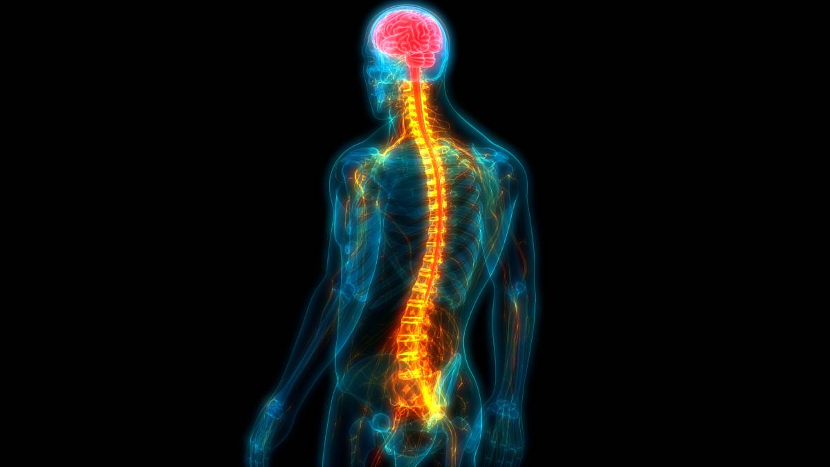
While pelvic misalignment is common in men, women are particularly vulnerable due to their larger and more mobile pelvises. A report funded by the National Institutes of Health found that nearly 24 percent of women in the U.S. are affected by one or more pelvic floor disorders and that the frequency of pelvic floor disorders increases with age (U.S. Department of Health and Human Services 2015). The imbalance is especially prevalent during pregnancy as a woman’s body releases hormones that relax ligaments to create space for the developing fetus. The increased width of the pelvis often destabilizes the spine and, due to the added pressure from the weight of the child, shifts the spine out of alignment. A 2019 study in which pelvic measurements were obtained from 201 women at 12, 24, 30, and 36 weeks of pregnancy, and 1 month after childbirth found significant differences in the measurements over time (Morino 2019). The anterior and posterior width of the pelvis—the distance between the bilateral anterior/posterior superior iliac spines—became significantly wider as pregnancy progressed. Furthermore, the degree of pelvic tilt of the anterior pelvis increased during pregnancy. The anterior width of the pelvis was not recovered even at one month post-childbirth.
Knee pain is a common side effect of pelvic imbalance. A 2010 study examined the relationship between pelvis malposition and knee pain in 100 endurance runners, 50 with knee pain and 50 without knee pain, and concluded that there is a correlation between one-sided pelvic mispositioning and knee pain during activities such as endurance running (Siegele 2010).
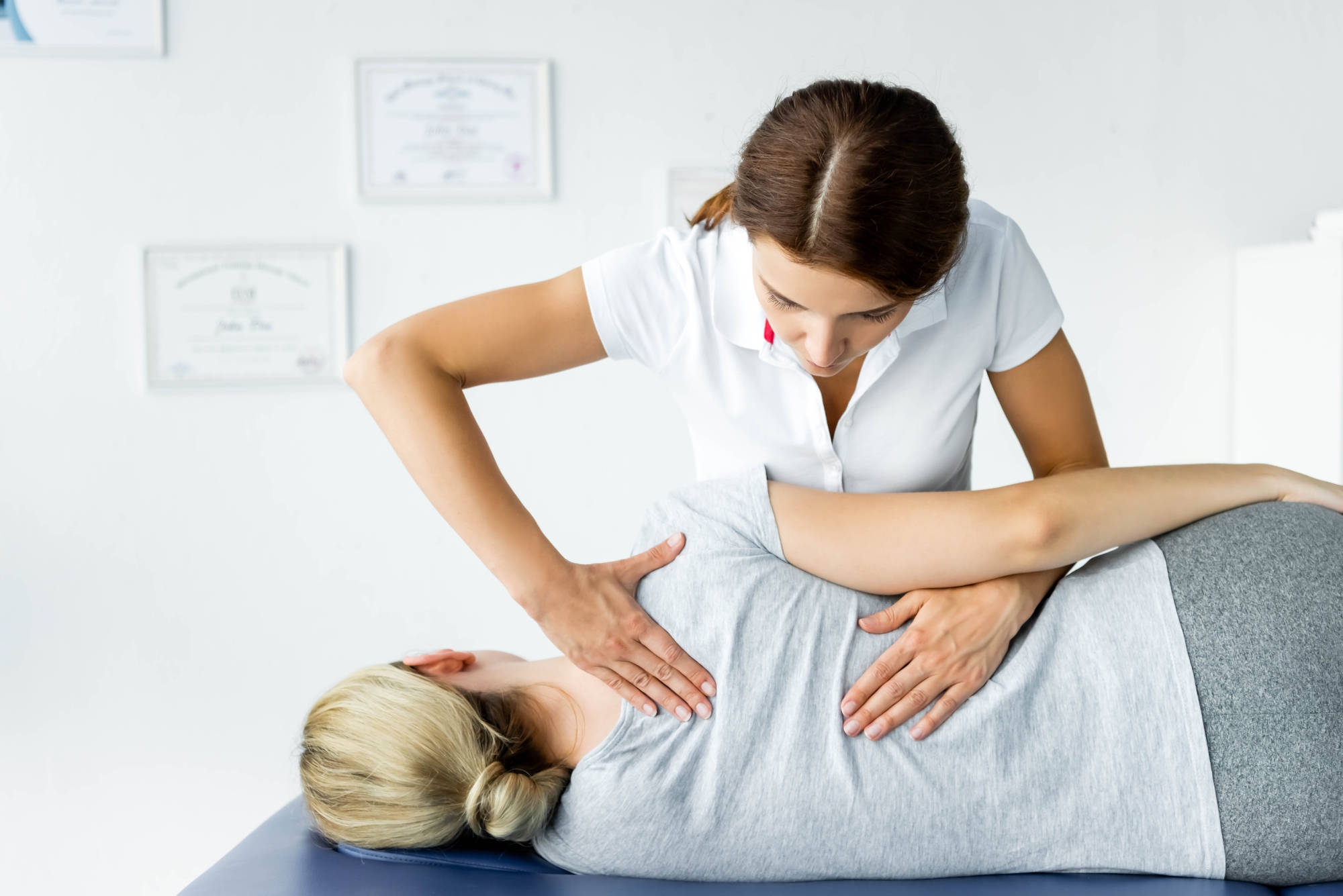
Understanding the causes of pelvic misalignment and devising a treatment plan may help avert knee complications or alleviate pain. One case report describes successful chiropractic treatment of a female patient’s knee pain stemming from pelvic imbalance (Bucek 2019). The patient complained of ongoing anteromedial knee pain when walking and a persistent feeling of her knee “giving out” after walking no more than a half mile. The patient was administered five treatments over the course of ten days, and in each treatment received chiropractic manipulation of the sacroiliac joint, kinesiology taping, and gluteus medius exercises to improve muscle strength. Following the treatments, she reported a resolution of her knee issues and experienced no pain while walking or performing other activities (Bucek 2019).
As described, conservative measures to correct imbalance and resolve knee pain may be effective for patients experiencing knee complications stemming from pelvic misalignment. Chiropractic treatments may help increase muscle strength and flexibility as well as ease the tension on supportive tissues that are strained from misaligned or stiff pelvic joints.
References
Bucek, D. (2019). Reduction of Knee Pain in a 45-Year-Old Woman After Pelvic Manipulation and Kinesiology Taping: A Case Report. Journal of Chiropractic Medicine, 18(3), 236-241. https://doi.org/10.1016/j.jcm.2019.07.006.
First Digital Solutions. (2016, October 14). Pelvic Imbalance. Chelsea Osteopathic Clinic. https://chelseaosteopaths.co.uk/pelvic-imbalance/.
Karp Rehabilitation. (2018, February 20). Understanding Pelvic Injuries & How to Treat Them. Karp Rehabilitation. http://karprehab.com/pelvic-injuries/.
Morino, S., et al. (2019). Pelvic alignment changes during the perinatal period. PloS one, 14(10), e0223776. https://doi.org/10.1371/journal.pone.0223776.
Siegele, J., et al. (2010). Relation between pelvis malposition and functional knee pain by long distance running. Sportverletzung Sportschaden, 24(3), 144–149. https://doi.org/10.1055/s-0029-1245638.
U.S. Department of Health and Human Services. (2015, September 28). Roughly One Quarter of U.S. Women Affected by Pelvic Floor Disorders. National Institutes of Health. https://www.nih.gov/news-events/news-releases/roughly-one-quarter-us-women-affected-pelvic-floor-disorders.