Common Gym Injuries That Chiropractors Treat
Chiropractic care is an increasingly popular treatment option for individuals who are involved in sports, fitness, and recreational activities. It focuses on the diagnosis, treatment, and prevention of musculoskeletal injuries, and is known for its effectiveness in addressing various ailments. Gym enthusiasts often face a myriad of injuries that can be debilitating if left untreated. Here are some of the most common gym injuries that chiropractors treat:
Sprains and strains: Overstretching or tearing of ligaments (sprains) and muscles or tendons (strains) are among the most common injuries that can occur during gym workouts. These injuries often result from overexertion, poor form, or inadequate warm-up. Chiropractic care can help alleviate pain and inflammation while promoting tissue repair and restoring joint mobility.
Lower back pain: Improper lifting technique, poor posture, and muscle imbalances can contribute to lower back pain, which is a common complaint among gym-goers. Chiropractors can provide spinal adjustments and soft tissue treatments to relieve pain and discomfort, as well as recommend exercises to strengthen core muscles and improve posture.
Shoulder injuries: The shoulder joint is highly susceptible to injury during gym workouts due to its extensive range of motion. Rotator cuff injuries, impingement syndrome, and dislocations are common problems that can be addressed by chiropractors. Treatment may include spinal and extremity adjustments, myofascial release, and rehabilitative exercises to restore function and prevent further damage.
Runner’s knee: Also known as patellofemoral pain syndrome, runner’s knee is a common overuse injury among gym enthusiasts who engage in high-impact activities like running, jumping, or plyometrics. Chiropractic care can help correct biomechanical imbalances, improve joint function, and provide pain relief through adjustments, soft tissue therapies, and tailored exercise programs.
Tennis elbow: Characterized by pain in the outer part of the elbow, tennis elbow is a result of overuse and repetitive stress on the tendons that connect the forearm muscles to the elbow. Chiropractors can employ a combination of joint manipulation, soft tissue techniques, and rehabilitative exercises to alleviate pain and promote healing.
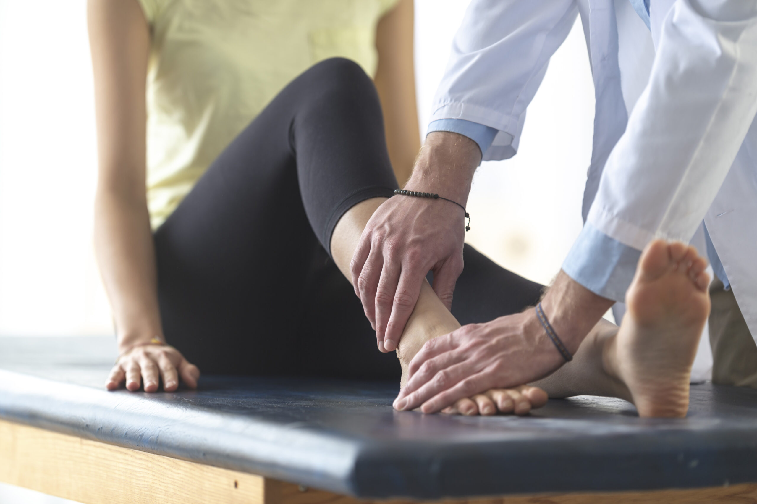
Achilles tendinitis: This condition involves inflammation and irritation of the Achilles tendon, often due to overuse or a sudden increase in physical activity. Chiropractic care can be beneficial in addressing the root cause of the problem, providing pain relief, and promoting a gradual return to activity through manual therapies and exercise recommendations.
Shin splints: Medial tibial stress syndrome, or shin splints, is a common injury among gym enthusiasts who engage in high-impact activities. Chiropractors can help alleviate pain and inflammation by addressing biomechanical imbalances, providing soft tissue therapies, and recommending appropriate strengthening and stretching exercises.
In conclusion, chiropractic care can be a valuable resource for gym enthusiasts who are suffering from various injuries. By addressing the underlying causes of these injuries and providing targeted treatment, chiropractors can help individuals return to their favorite activities more quickly and safely. Remember to consult with a qualified chiropractor if you’re experiencing any of the aforementioned issues or other gym-related injuries.