Vertigo is an abnormal sensation of motion or loss of balance and can be either chronic or intermittent. Brief sensations of vertigo may arise for a wide variety of reasons, such as sitting up quickly or playing three-dimensional video games. More persistent forms of vertigo usually arise due to dysfunction of the inner ear, which is responsible for our sense of balance and positioning. Cold viruses, head trauma, and Meniere’s disease are all conditions that affect the inner ear and can cause a sensation of unsteadiness (The University of Iowa 2018). Musculoskeletal conditions, including nerve damage to the legs, muscle weakness, and joint instability, may also give rise to vertigo and cause difficulties with movement. Finally, certain conditions such as Parkinson’s disease, depression, and impaired vision can be catalysts for vertigo (Mayo Foundation for Medical Education and Research 2020).
It is extremely rare for young children to be affected by vertigo, and it almost always occurs in adulthood. Approximately one in 20 adults experience vertigo each year, and the majority of people with the condition find that it severely impairs their daily activities and ability to work (van Vugt 2017). Furthermore, falls are the leading cause of injury among senior citizens and can cause severe trauma such as fractures. As falls are largely caused by impairments in balance, the potential consequences of vertigo become more severe with age.
If vertigo is accompanied by tinnitus, ear pressure, or hearing loss, the inner ear may be the source of the condition. An otolaryngologist, a specialist in ear disorders, may examine the onset, duration, and severity of ear discomfort experienced. Other symptoms that commonly accompany ear disorders include nausea and vomiting, as well as lowered heart rate (The University of Iowa 2018). Positional vertigo, a form of vertigo in which symptoms change depending on the position of the head, is common and primarily originates in the inner ear.
Vertigo is usually treatable with physical therapy, medication, and/or surgery (The University of Iowa 2018). Neurologists typically order tests and scans to determine the cause of the condition before developing a treatment plan. Symptoms of vertigo caused by more serious structural problems may require surgery, while in some cases, balance exercises and lifestyle changes alone can help manage symptoms. Other conservative treatments, such as restricting foods and drinks that cause migraines or impair the senses (e.g. alcohol and coffee), may be recommended by a medical professional.
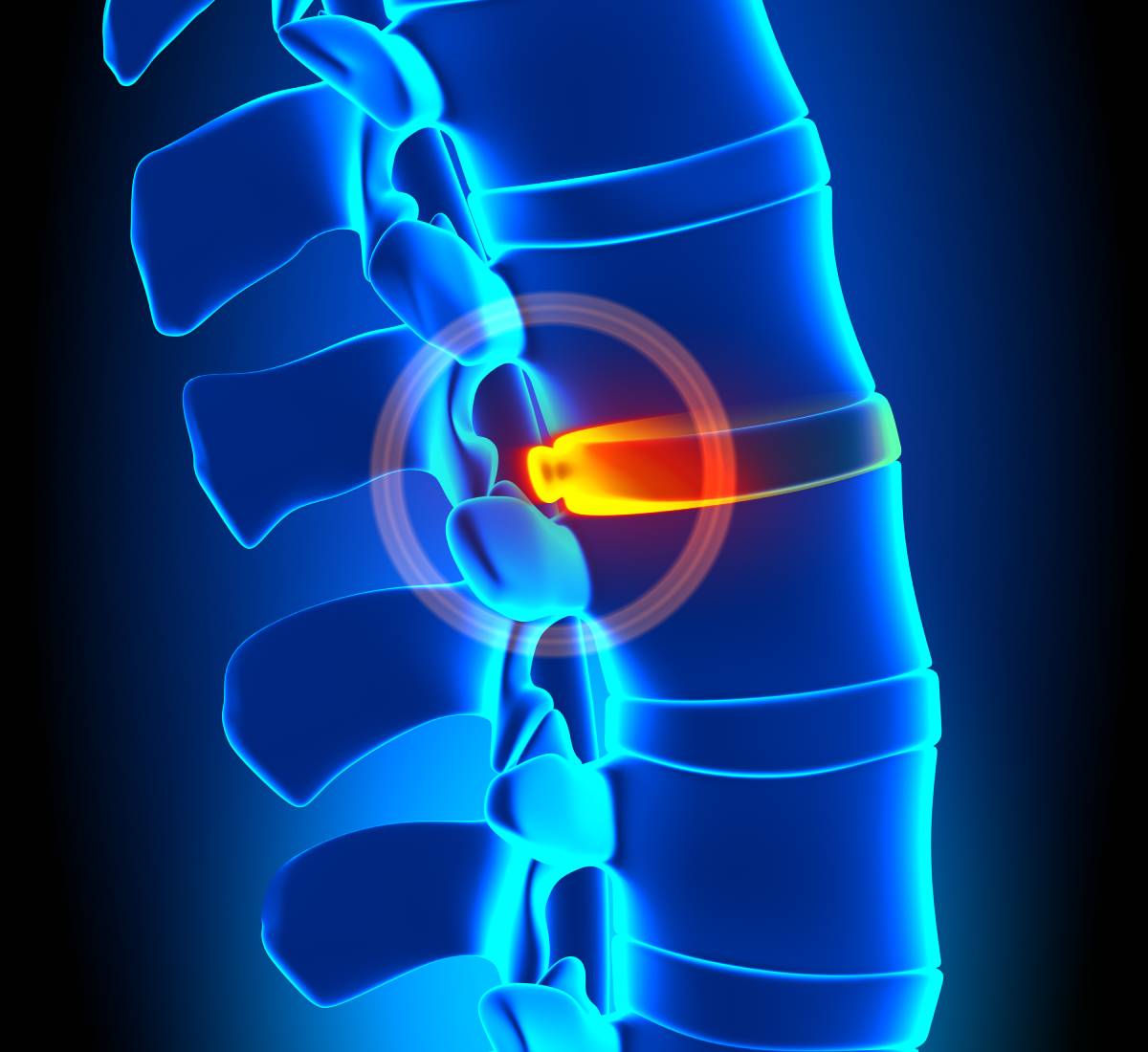
Chiropractic care is another effective form of treatment for vertigo that uses hands-on manipulation to help patients improve their balance and coordination. According to a 2010 survey conducted by the National Board of Chiropractic Examiners, chiropractors report seeing, on average, between one and three patients per month for concerns about dizziness (Ndetan 2016). Some studies suggest that chiropractic manipulations targeting the cervical spine may be helpful in treating balance disorders, such as vertigo, by re-positioning the neck in its optimal location and bringing the body back to equilibrium.
A 2009 study examined the effects of spinal manipulation and manual therapy on dizziness and balance at a chiropractic college health center and a senior fitness center (Strunk 2009). A group of 19 adults, aged 40 years or older with a median age of 70, completed the study. All patients were treated by either a clinician or a chiropractic student intern twice per week, each session lasting 15-20 minutes, during an 8-week intervention period. The Dizziness Handicap Inventory, the Short Form Berg Balance Scale (SF-BBS), and the Neck Disability Index were used to measure the effects of the treatments. A large difference in the SF-BBS before and after the intervention period was measured in most patients, demonstrating an improvement in balance. Some patients also showed reduced dizziness and neck pain at the conclusion of the study.
References
Mayo Foundation for Medical Education and Research. (2020). Balance Problems. Mayo Clinic. https://www.mayoclinic.org/diseases-conditions/balance-problems/symptoms-causes/syc-20350474#.
Ndetan, H., et al. (2016). The Role of Chiropractic Care in the Treatment of Dizziness or Balance Disorders: Analysis of National Health Interview Survey Data. J Evid Based Complement Altern Med, 21:138–142. doi:10.1177/2156587215604974.
Strunk, R., et al. (2009). Effects of Chiropractic Care on Dizziness, Neck Pain, and Balance: A Single-Group, Preexperimental, Feasibility Study. Journal of Chiropractic Medicine, 8(4), 156–164. doi:10.1016/j.jcm.2009.08.002.
The University of Iowa. (2018). Vertigo: Frequently Asked Questions. The University of Iowa Hospitals & Clinics. https://uihc.org/health-topics/vertigo-frequently-asked-questions.
van Vugt, V., et al. (2017). Chronic Vertigo: Treat with Exercise, Not Drugs. BMJ Publishing Group, 358. doi:10.1136/bmj.j3727.